


This article is drawn in particular from the SENORIF 2025-2026 guidelines.

Does your biopsy report mention lobular carcinoma in situ?
Bring it with your images: Dr Jérémie Zeitoun, breast cancer surgeon, reviews the file and tells you whether to operate or to monitor, with the reason for that choice.
High-risk lesion, not a cancer: what that means
The lobule, where the lesion arises
The breast contains ducts, which carry the milk, and lobules, the small glands that produce it. Each lobule is made up of tiny cavities, the acini.
In lobular carcinoma in situ, abnormal cells, which no longer adhere to one another, fill and distend these acini. “In situ” means “in place”: the cells stay inside the lobule and have not invaded the surrounding tissue.
Classic lobular carcinoma in situ is therefore not a breast cancer. It is one of the high-risk lesions of the breast, also called “B3 lesions” or “borderline lesions”, because they are neither entirely benign nor a cancer.
It does not necessarily turn into a cancer: it signals a higher risk of breast cancer, in either breast.
It is most often discovered by chance, when exploring microcalcifications, the small calcium deposits seen on the mammogram (see breast microcalcifications). It accounts for 0.5 to 4% of biopsies of benign breast lesions, according to the 2022 recommendations of the French National College of Gynaecologists and Obstetricians.
The three forms of lobular carcinoma in situ
The report must state the form, because the decision depends on it.
The classic form
It is by far the most common: a high-risk lesion, not a cancer. In a woman who has it, the risk of breast cancer is multiplied by 8 to 10 compared with a woman without this lesion.
The French National College of Gynaecologists and Obstetricians estimated this risk, in 2022, at 1 to 2% per year in women. It often affects several areas of the same breast and, in 30 to 67% of cases, both breasts.
The pleomorphic form
The cell nuclei are at least four times larger than that of a lymphocyte, a white blood cell taken as a point of comparison. The lesion resembles a high-grade ductal carcinoma in situ. It is rarer.
The florid form
The lobule is very distended, almost like a mass, with little or no tissue between the cavities. It used to be called “with necrosis”. Necrosis, an area of dead cells at the centre of the lesion, can accompany the florid form as well as the pleomorphic form.
These two forms are generally limited to a single area, and can coexist with an invasive lobular carcinoma or with the classic form. The guidelines consider them “aggressive” and manage them “in the same way as ductal carcinoma in situ”. They are not simple high-risk lesions: their management differs from that of the classic form.
After the biopsy: why surgery is most often performed
What the biopsy does not always show
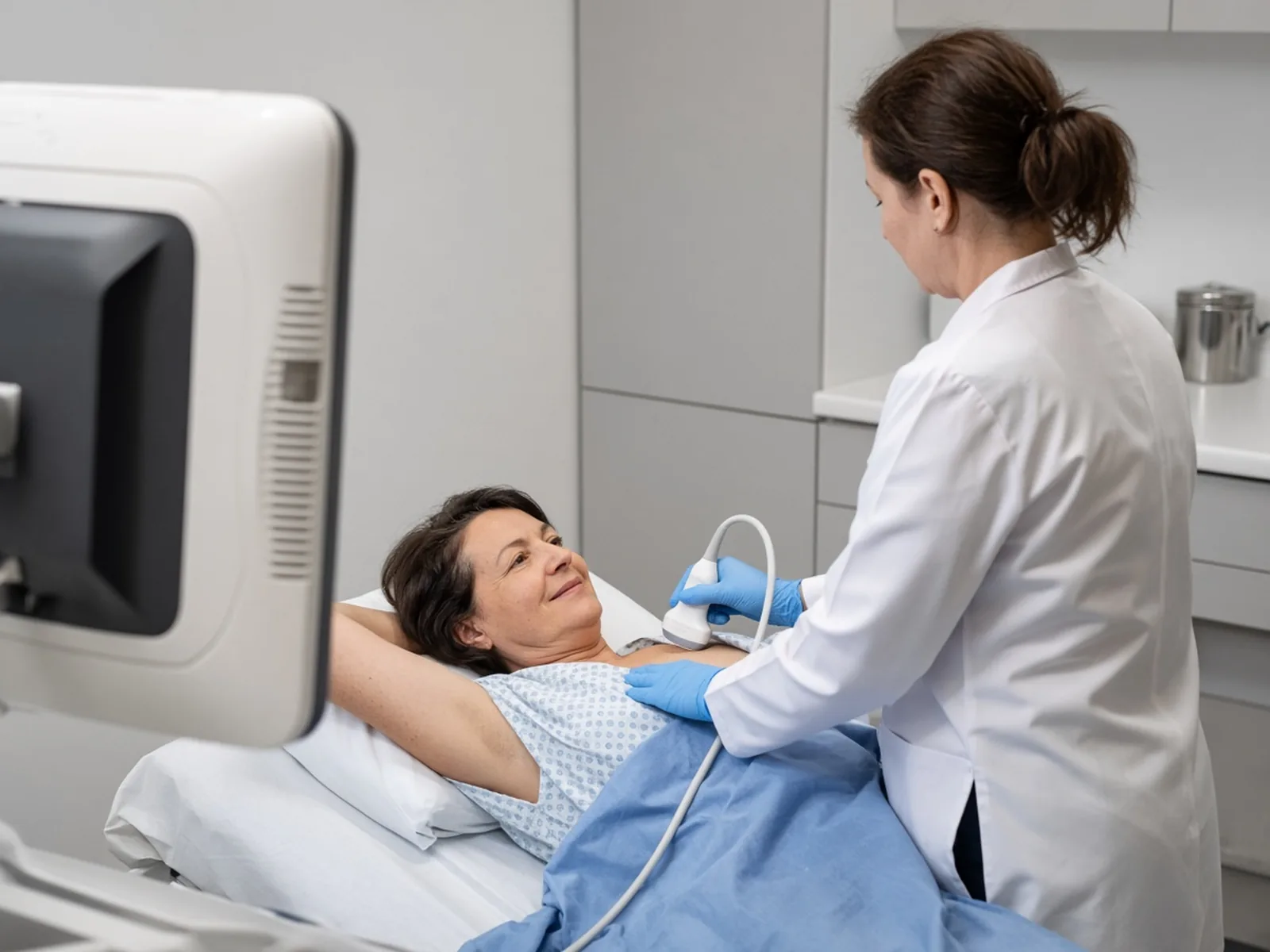
The biopsy samples a fragment of the suspicious area, not the whole area. Next to the lesion, there may be a cancer that the sample did not reach: this is underestimation.
That is why, when the biopsy shows lobular carcinoma in situ, surgery is most often performed: analysis of the area removed makes it possible to be sure that there is no cancer next to it.
For the classic form, with no other associated lesion and when the images match the biopsy result, the proportion of cancers discovered at surgery is low: less than 5% according to the 2023 European guidelines on B3 lesions, 0 to 4% according to the American Society of Breast Surgeons. It rises markedly for the pleomorphic and florid forms: according to American series, often from a single centre, 25 to 60% for the pleomorphic form and 30 to 40% for the florid form.
These figures are orders of magnitude, not your personal risk.
The check after every biopsy
After every biopsy, whatever the result, we check that the result matches what the images show: this is radiological-pathological concordance (radiological for the imaging, pathological for the examination under the microscope). This check is systematic. In the event of discordance, new samples or surgery are offered: see discordance between imaging and biopsy.
Core needle biopsy or vacuum-assisted biopsy
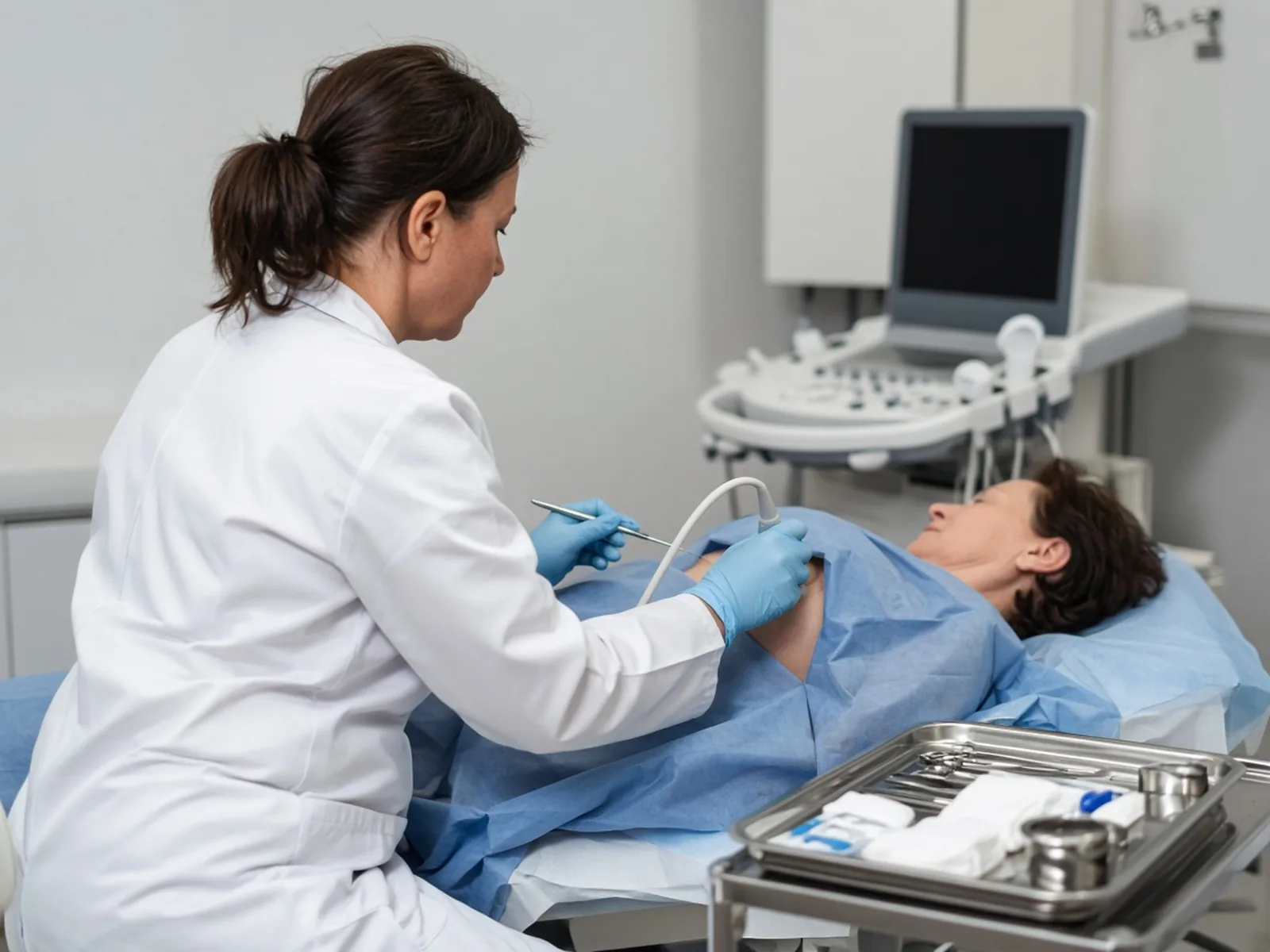
The core needle biopsy takes fine cores with a needle. The vacuum-assisted biopsy, performed by the radiologist under image guidance, takes more tissue and can sometimes remove the whole visible area (see breast biopsy). According to the guidelines, for the classic form:
- diagnosis on core needle biopsy: a vacuum-assisted excision is offered to remove the area;
- diagnosis on vacuum-assisted biopsy: surgical excision in the event of discordance or if a lesion remains visible on the images; otherwise, follow-up by imaging is possible, under the conditions described below. Most often, Dr Zeitoun operates.
For the pleomorphic and florid forms, management is that of ductal carcinoma in situ: surgical excision.
The biopsy, the ultrasound and the mammogram are performed by the radiologist, who then refers you to Dr Zeitoun with the reports (see mammogram, breast ultrasound and breast magnetic resonance imaging (MRI)).
Surgery or surveillance: the decision guide
| Your situation | Course of action proposed |
|---|---|
| Classic form, discovered on core needle biopsy | Vacuum-assisted excision, or surgical excision |
| Classic form, discovered on vacuum-assisted biopsy | Most often, surgical excision. Surveillance is the exception, discussed case by case in a multidisciplinary team meeting and with you |
| Classic form with discordance, lesion remaining on the images or another associated atypical lesion | Surgical excision, then surveillance |
| Pleomorphic or florid form | Multidisciplinary team meeting, then surgical excision |
| Classic form, margins involved after the operation | No further operation, no additional treatment, no radiotherapy |
| Pleomorphic or florid form, margins involved | Further excision |
| Pleomorphic form or form with necrosis, after excision | Radiotherapy to be discussed |

Are you hesitating between surgery and surveillance?
It is a shared decision, made knowing the form of the lobular carcinoma in situ and your images: bring your questions, Dr Zeitoun presents both options and their consequences.
Surveillance without surgery: the exception
For the classic form, surgery is most often performed. The guidelines nevertheless allow follow-up by imaging, without surgery, only if all the following conditions are met:
- the target has completely disappeared after the vacuum-assisted biopsy;
- the biopsy and the imaging are concordant;
- the cluster of microcalcifications measured less than 15 mm, and there was no more than one cluster;
- no mass, no architectural distortion, no ultrasound correlate;
- no lesion causing symptoms;
- no personal or genetic risk factor;
- an elderly patient who accepts close surveillance.
Even when these conditions are met, surveillance remains the exception: it is decided case by case, in a multidisciplinary team meeting, where several doctors review your file, and with you.
Pleomorphic or florid forms: what the guidelines say
The guidelines manage them “in the same way as ductal carcinoma in situ”. Before surgery for breast cancer, they recommend a multidisciplinary team meeting, where at least three expert doctors, including a surgeon, a radiation oncologist and a medical oncologist, examine the file together.
Dr Zeitoun therefore presents these files at a multidisciplinary team meeting before the excision.
The pathology report on the specimen must then state the form (florid, “with or without necrosis”, or pleomorphic), the size of the lesion and its distance from the margins. The guidelines note that the ideal margin for these forms is not known.
If an operation is proposed
Excision of a lesion that cannot be felt
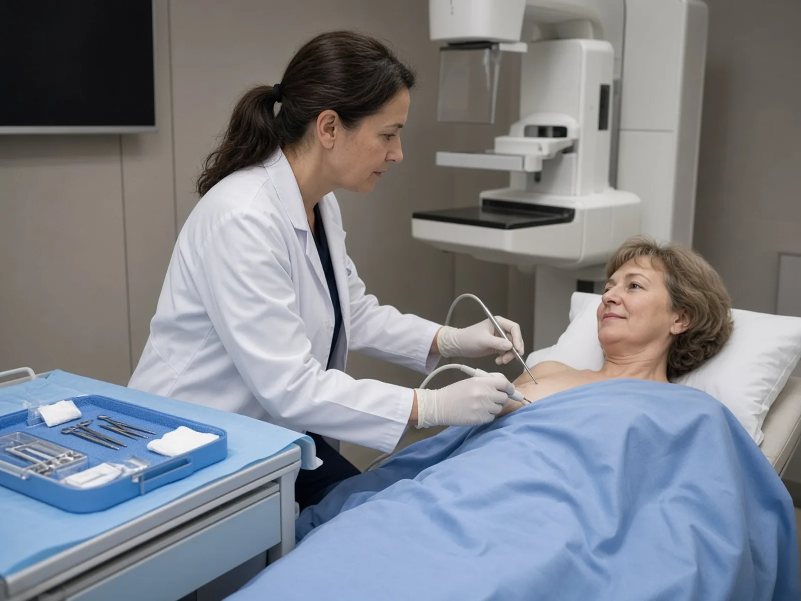
Lobular carcinoma in situ generally cannot be felt. The radiologist therefore places a hookwire before the procedure: a fine metal wire, placed under local anaesthesia on the morning of the operation or the day before, which shows the surgeon where to operate (see preoperative localisation).
The marked area is then removed by a conservative excision (see lumpectomy): the breast is preserved. The specimen is X-rayed during the procedure to check that the lesion has indeed been removed.
The excision is done as day surgery, at Clinique Hartmann: you go home the same day. See surgery for high-risk lesions of the breast, preparing for your operation and postoperative recovery.
The margins and the report
The specimen removed is analysed under the microscope. The margins are the edges of the specimen: we look at whether abnormal cells touch them.
- Classic form: if the margins are involved, there is no re-excision. It is not a cancer but a high-risk lesion: a further operation is not recommended. There is no additional treatment. Surveillance of both breasts is enough.
- Pleomorphic or florid form: if the margins are involved, a further excision is recommended.
Analysis of the specimen can also reveal a cancer that the biopsy had not seen, which then changes the management. See understanding your pathology report and margins and re-excision after lumpectomy.
Radiotherapy
Radiotherapy is a treatment with rays, delivered to the breast after the operation. For the classic form, treated by conservative surgery, there is no indication for radiotherapy: no additional treatment is necessary. Radiotherapy is indicated only for the pleomorphic forms or forms with necrosis, as necrosis can accompany the florid form as well as the pleomorphic form.
Surveillance after lobular carcinoma in situ
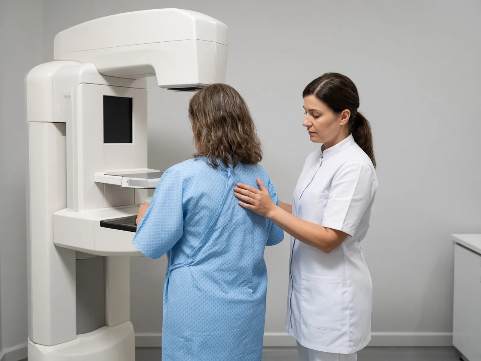
Whether or not surgery has been performed, the lesion requires surveillance of both breasts. After lobular carcinoma in situ, the guidelines provide for:
- a clinical breast examination every year;
- a two-view mammogram every year for 10 years, then every 2 years;
- an ultrasound, as an option;
- no systematic breast MRI.
The BI-RADS classification (Breast Imaging-Reporting and Data System), which grades each breast imaging examination, says whether further steps are needed: see understanding BI-RADS. If your breasts are dense, see dense breasts: what surveillance.
If you have a family or genetic risk, risk assessment is detailed in the risk assessment tools and in risk of breast and ovarian cancer.
Why there is no mastectomy
Lobular carcinoma in situ does not justify removing the breast. The French guidelines state: “no indication for prophylactic mastectomy” for lobular carcinoma in situ.
Those of the French National College of Gynaecologists and Obstetricians of 2022 point the same way: in the absence of a demonstrated benefit on survival and quality of life, it is not recommended to perform a risk-reducing mastectomy for a high-risk lesion of the breast (strong recommendation).
For the classic form, it is a high-risk lesion, not a cancer: the answer is regular surveillance of both breasts, and not removal.
The rare exception
The French National College of Gynaecologists and Obstetricians of 2022 provides for a single exception, a weak recommendation: pleomorphic or florid lobular carcinomas in situ, multicentric or diffuse, associated with complex imaging or other risk factors, in a young woman (under 40). In this case, the file is discussed in a multidisciplinary team meeting.
Preventive surgery can also be discussed in other situations, such as a BRCA1 or BRCA2 gene mutation, always in a multidisciplinary team meeting. See preventive mastectomy, bilateral preventive mastectomy, reconstruction after preventive mastectomy and preventive surgery in the case of BRCA.

Would you like a second opinion before deciding?
Excision, surveillance or a simple question about your report: Dr Zeitoun reviews your reports and your images, and tells you what he would do in your place, with no obligation.
What you should expect from the consultation

What to bring
- The biopsy report, with the form of the lobular carcinoma in situ if it is stated.
- The mammogram and ultrasound reports and, if there is one, the MRI report, and the images on disc or online.
- The list of your personal and family history of breast cancer or ovarian cancer, and your genetic results if any.
See preparing for your first consultation and, for an additional opinion, breast cancer second opinion.
Where and with whom
Consultations at the practice at 241 rue du Faubourg Saint-Honoré (Paris 8th) and at Clinique Hartmann (Neuilly-sur-Seine). Surgery takes place at Clinique Hartmann.
Patients from abroad will find the arrangements in international patients. To learn about Dr Zeitoun’s background, see his background and breast cancer surgeon in Paris.
Frequently asked questions
Is lobular carcinoma in situ (LCIS) a cancer?
In its classic form, no: it is a high-risk lesion, not a cancer. The cells stay in the lobules and the lesion mainly signals a higher risk of breast cancer in both breasts. The pleomorphic and florid forms are closer to a cancer: they are managed like ductal carcinoma in situ.
Should lobular carcinoma in situ always be operated on?
No, but most often yes: when the biopsy shows lobular carcinoma in situ, it is most often operated on. For the pleomorphic and florid forms, surgical excision is the course of action, after a multidisciplinary team meeting. For the classic form, surveillance is the exception: it is possible only under precise conditions, and is decided case by case in a multidisciplinary team meeting and with you.
What does “radiological-pathological concordance” mean?
After every biopsy, we check that the biopsy result matches what the images show: radiological for the imaging, pathological for the examination under the microscope. This check is systematic. In the event of discordance, new samples or surgery are offered. See discordance between imaging and biopsy.
The margins are not clear: is a second operation needed?
For the classic form, no: there is no re-excision, because it is not a cancer but a high-risk lesion, and no additional treatment is necessary. For the pleomorphic and florid forms, a further excision is recommended. See margins and re-excision after lumpectomy.
Is radiotherapy needed after the operation?
Not for the classic form: no additional treatment is necessary. It is indicated only for the pleomorphic forms or forms with necrosis. See radiotherapy.
Can lobular carcinoma in situ affect the other breast?
Yes. The classic form often affects several areas of the same breast and, in 30 to 67% of cases, both breasts. That is why surveillance covers both breasts.
How does surveillance work, and is an MRI needed?
Clinical examination every year, two-view mammogram every year for 10 years, then every 2 years. Magnetic resonance imaging (MRI) is not systematic. See mammogram.
Is mastectomy recommended?
No, as a rule. The French guidelines do not provide for preventive mastectomy for lobular carcinoma in situ. The only exception, a weak recommendation, concerns a multicentric or diffuse pleomorphic or florid form, with complex imaging or other risk factors, in a woman under 40. See preventive mastectomy.
Is the operation done as day surgery?
Yes. The hookwire is placed on the morning of the procedure or the day before, the excision takes place at Clinique Hartmann and you go home the same day. See postoperative recovery.
What are Dr Zeitoun’s fees?
Dr Zeitoun practises in Sector 2 (non-OPTAM) and charges fees above the French Social Security rate for all consultations and procedures. Most international patients are not covered by French Social Security. A detailed quote is given during the consultation, before any procedure. If you are covered, the Assurance Maladie reimburses on the basis of the Social Security rate, and your health insurance may cover all or part of the additional fees, depending on your policy. See quote, stay and cover.
Sources
- Cancers et pathologies du sein : attitudes diagnostiques et thérapeutiques, protocoles de traitement, SENORIF 2025-2026. Table of high-risk lesions (classic lobular carcinoma in situ ×8-10), management according to the type of biopsy, conditions for surveillance, margins, no indication for prophylactic mastectomy, follow-up, content of the pathology report, use of the multidisciplinary team meeting.
- Mathelin C. et al., Collège national des gynécologues et obstétriciens français : Indications des mastectomies de réduction de risque en dehors du risque avéré de prédisposition héréditaire, recommandations pour la pratique clinique, 2022. Gynécologie Obstétrique Fertilité & Sénologie, 2022, 50, 107-120. World Health Organization 2019 definition, frequency, subtypes, risk, no benefit of risk-reducing mastectomy, exception for the pleomorphic or florid forms.
- World Health Organization: WHO Classification of Tumours, Breast Tumours, 5th edition, 2019; Tan P.H. et al., The 2019 WHO classification of tumours of the breast. Histopathology, 2020, 77, 181-185. Three forms (classic, pleomorphic, florid).
- Rubio I.T. et al., European guidelines for the diagnosis, treatment and follow-up of breast lesions with uncertain malignant potential (B3 lesions), developed jointly by the European Society of Breast Cancer Specialists (EUSOMA), the European Society of Breast Imaging (EUSOBI), the European Society of Pathology (ESP) and the European Society of Surgical Oncology (ESSO). European Journal of Surgical Oncology, 2024, 50, 107292. Vacuum-assisted excision, surveillance in the event of concordance, underestimation below 5%.
- American Society of Breast Surgeons: Resource Guide, Surgical Management of Benign or High-Risk Lesions (archived document). Underestimation according to the form.
Article written by Dr Jérémie Zeitoun, breast and gynaecological cancer surgeon.