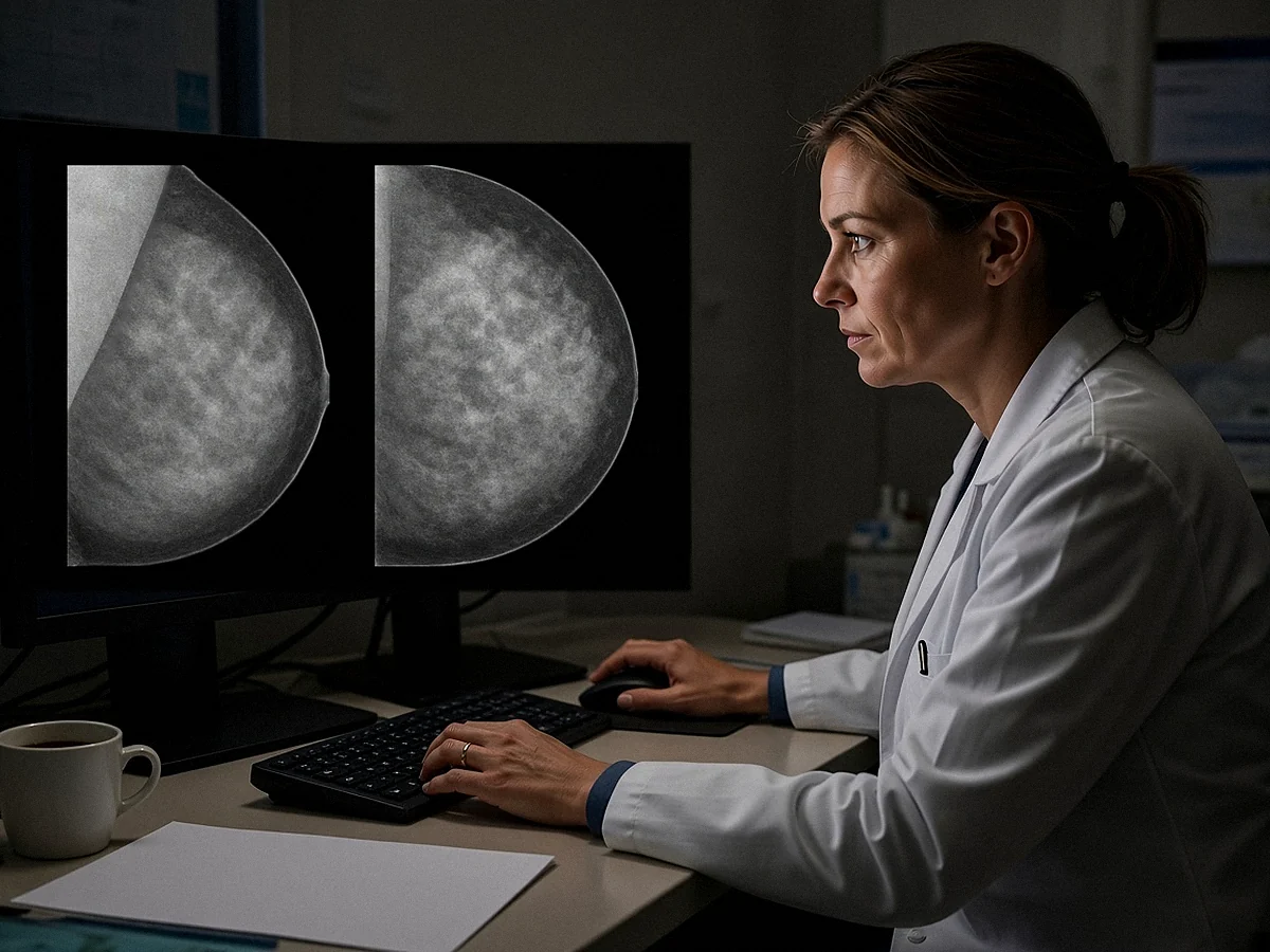


This article draws in particular on the SENORIF 2025-2026 guidelines.
Your biopsy is reassuring, but you are being told about discordance or surgery?
Bring the biopsy report and the images. Dr Jérémie Zeitoun, cancer and breast surgeon, sees patients at the practice at 241 rue du Faubourg Saint-Honoré (Paris 8th) and at Clinique Hartmann (Neuilly-sur-Seine). Surgery takes place at Clinique Hartmann.
An online consultation is possible. You can book an appointment on Doctolib or request a callback from the secretariat.
What radiologic-pathologic concordance means

A biopsy result is never read on its own. It is read alongside the image that led to the biopsy.
The radiologist first describes the abnormality and classifies it according to its level of suspicion. An image classified ACR 4 or ACR 5 leads to a sample being taken. This classification does not name a disease: it says how suspicious the image is.
Five categories
The sample is then analysed by the pathologist, the doctor who examines tissue under the microscope. The pathology report classifies the result into five categories:
| Category | What it means | What usually follows |
|---|---|---|
| B1 | Normal or non-diagnostic tissue | Check that the target was sampled; most often, a repeat biopsy if the image is suspicious |
| B2 | Benign lesion, for example a fibroadenoma | If the result is concordant, generally no surgery; if it is discordant, a repeat biopsy or excision |
| B3 | Lesion of uncertain malignant potential | Excision in most cases, by surgery or by vacuum-assisted biopsy depending on the lesion |
| B4 | Suspicious lesion, without certainty | Repeat biopsy or diagnostic excision; no treatment surgery on this result alone |
| B5 | Cancer, in situ or invasive | Work-up and treatment discussed at the multidisciplinary team meeting |
Comparing the two
One last step remains, and it is essential: comparing the two. The radiologist who performed the biopsy reviews the result, compares it with the images and states whether this result explains what was seen.
The radiologist records this in the report, with a proposal for what comes next. If in doubt, the radiologist discusses it with the pathologist.
Concordant or discordant
A result is concordant when it explains the image. An oval, regular mass, classified ACR 4A, which corresponds to a fibroadenoma under the microscope: the image and the result tell the same story.
A result is discordant when it does not explain it. The image was highly suspicious, or the sample did not reach its target, and the word “benign” is not enough to reassure. In this case, the result cannot be accepted as the final diagnosis.
The most common situations of discordance
The most common discordances are a highly suspicious image with a benign result, a mass that grows, a cluster of microcalcifications missing from the samples, an architectural distortion and a B1 result.
A highly suspicious image with a benign result
This is the most typical situation. The image is classified ACR 5: an irregular mass with spiculated margins, or an abnormality that has all the features of a cancer. The biopsy comes back B2, for example unremarkable fibrous tissue.
This fibrous tissue does not explain such a suspicious image. The likely explanation is then that the needle sampled the tissue next to the lesion, and not the lesion itself. Even if the report is reassuring, the diagnosis must be completed.
A mass that grows
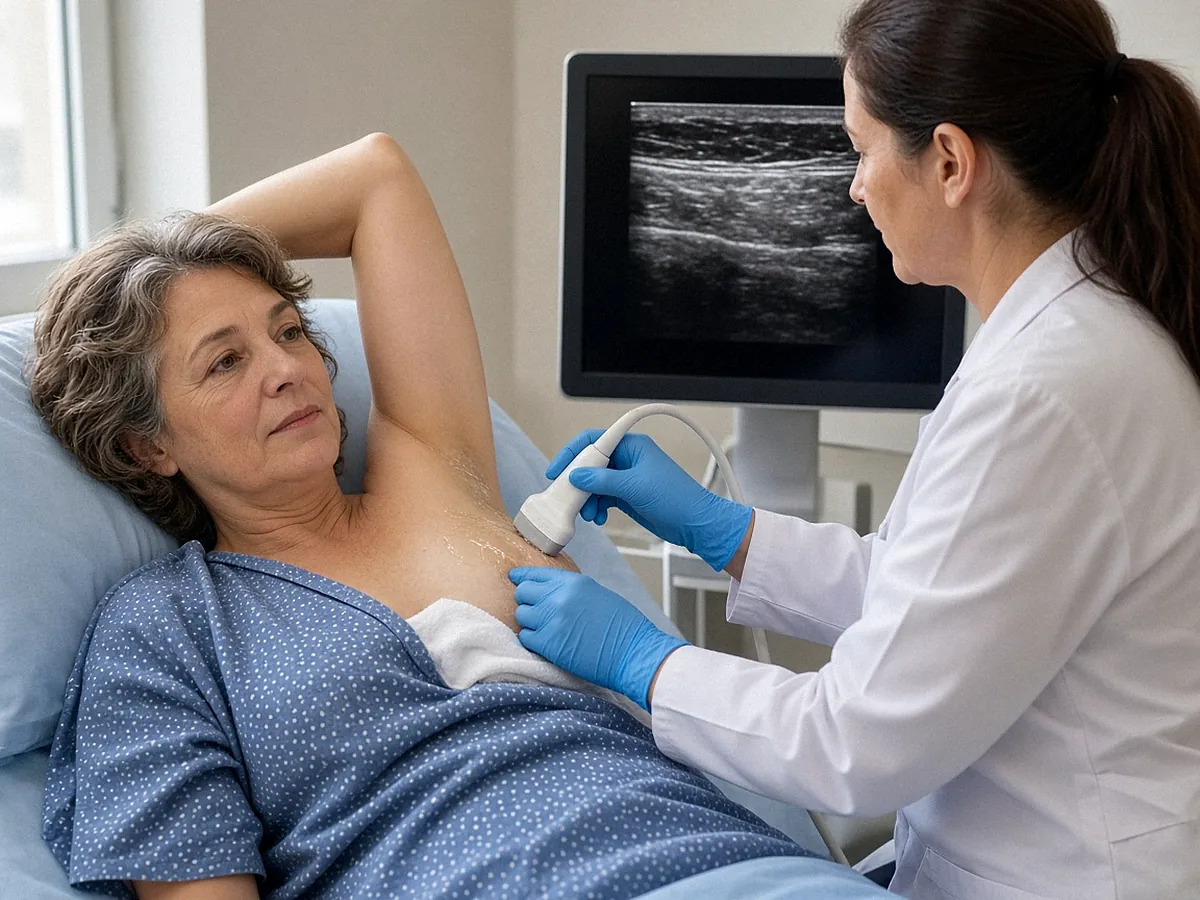
A mass has been biopsied, with a benign result. On the follow-up ultrasound, it has clearly increased in size, or its appearance has changed.
Some fibroadenomas grow. But rapid growth also raises the possibility of a phyllodes tumour, which is difficult to tell apart from a fibroadenoma on simple cores, or of a lesion that was poorly sampled. The lesion is then often re-sampled or removed.
A cluster of microcalcifications missing from the samples
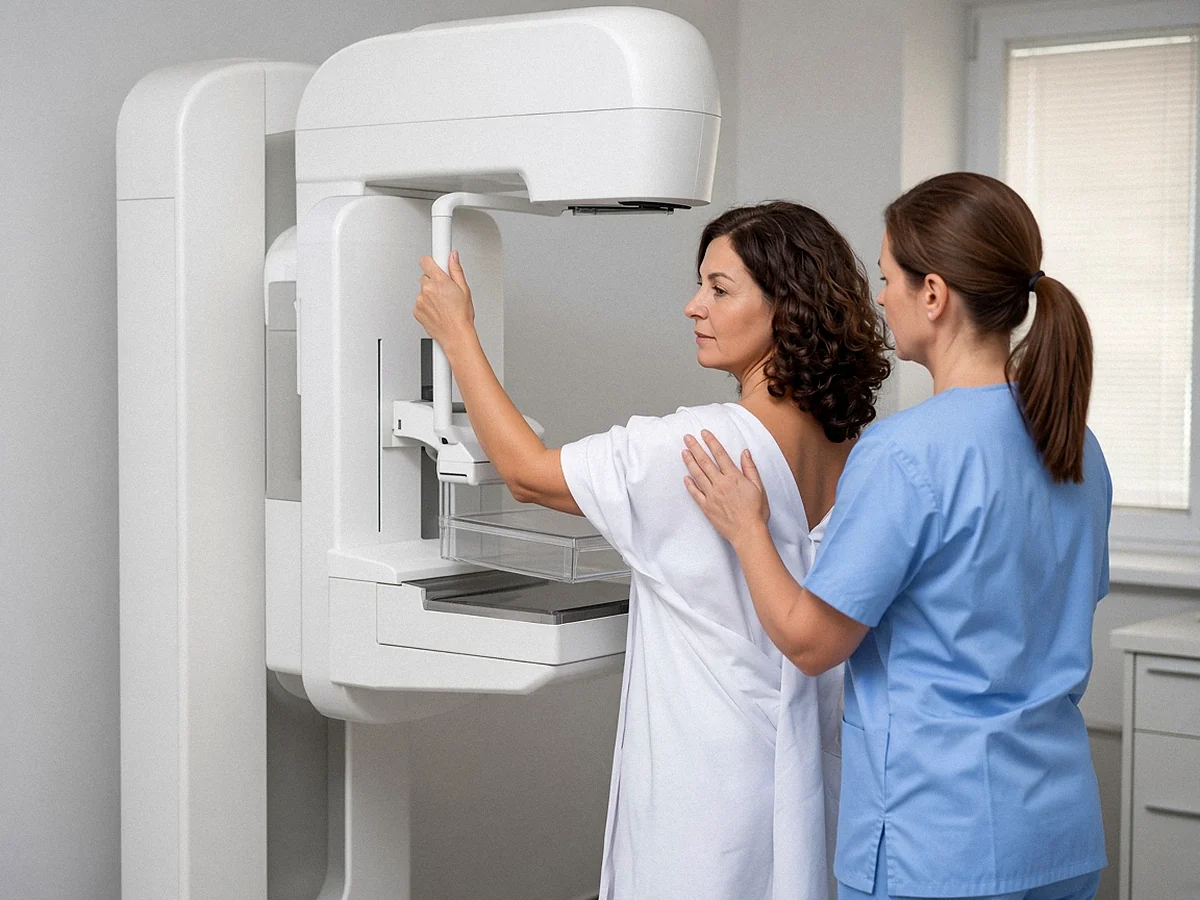
A cluster of microcalcifications is most often sampled by vacuum-assisted biopsy under stereotactic guidance, that is, guided by mammography. At the end of the procedure, the radiologist X-rays the samples taken to check that the microcalcifications are in them.
If these microcalcifications do not appear on the X-ray of the samples, or if the pathologist does not find them under the microscope, the result does not concern the right area. In this case, tissue without abnormality does not rule out a lesion.
An architectural distortion
An architectural distortion is an area where the gland appears pulled towards a point, without a distinct mass. It may correspond to a radial scar, to sclerosing adenosis, to an old scar, or to a cancer.
When the biopsy comes back with non-specific tissue that does not explain the distortion, the result is considered discordant. Sclerosing adenosis or a radial scar can, on the other hand, explain the image; it is for the radiologist to judge whether the result truly corresponds to what was seen.
A B1 result
A B1 result means that the sample contains normal tissue, or that it cannot be interpreted. With an image classified ACR 4 or ACR 5, it is not reassuring. It leads to checking that the right place was sampled, and most often to repeating the biopsy.
Why a biopsy may not be enough
A biopsy analyses a few fragments of the lesion, not the whole lesion. It therefore avoids surgery in the great majority of cases, but it may not represent the whole lesion.
Several reasons explain why a sample may not reach its target or may not fully represent it:
- a small, deep or mobile lesion, which moves away from the needle;
- dense breasts, in which the lesion is hard to make out;
- a heterogeneous lesion, partly benign and partly suspicious;
- an extensive cluster of microcalcifications, of which only part is sampled;
- a lesion visible only on MRI, for which there is no simple way of checking, during the procedure, that the right area has been sampled.
What the figures say
A systematic review published in 2026 brought together twenty-three studies on this subject. In series covering all biopsies, a discordant benign result concerns 1.2 to 5.3% of biopsies.
When these lesions are re-sampled or removed, a cancer is found in proportions that vary widely from one study to another. In the most representative series, this rate is around 20 to 40%. These are invasive cancers and ductal carcinomas in situ.
Most discordances are therefore benign, but the risk of cancer is too high to settle for surveillance.
What happens next
Faced with a discordant benign result, the guidelines propose two options: a repeat image-guided biopsy, or surgical excision. The choice depends on the image, the size of the lesion, its location and the type of sample already taken.
Reviewing the images
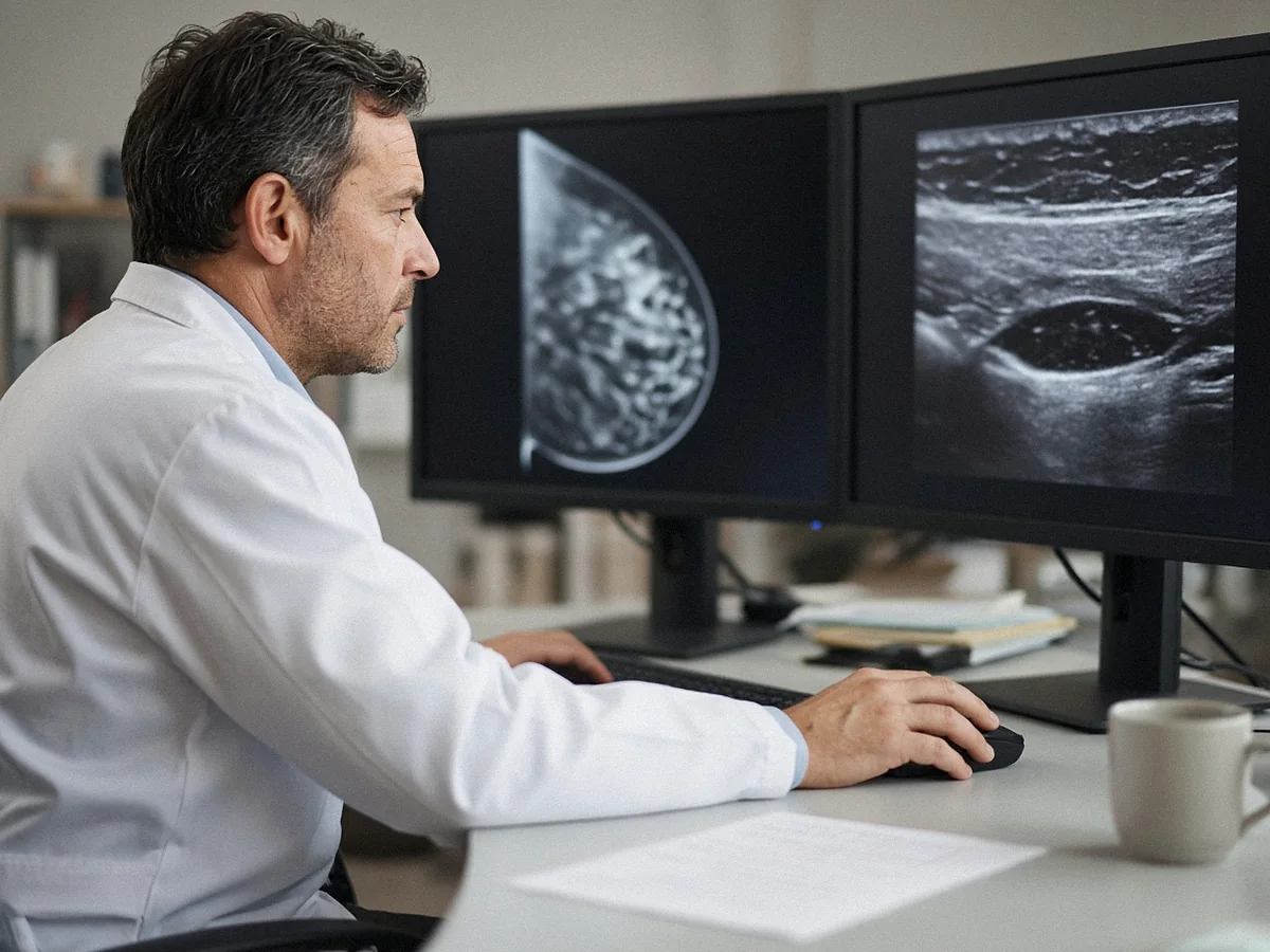
The first step is often a review. The radiologist looks at the images again, compares them with previous examinations, and may repeat a targeted ultrasound, sometimes called a “second look”.
This review can show whether the needle was really in the lesion. It can also lead to an MRI.
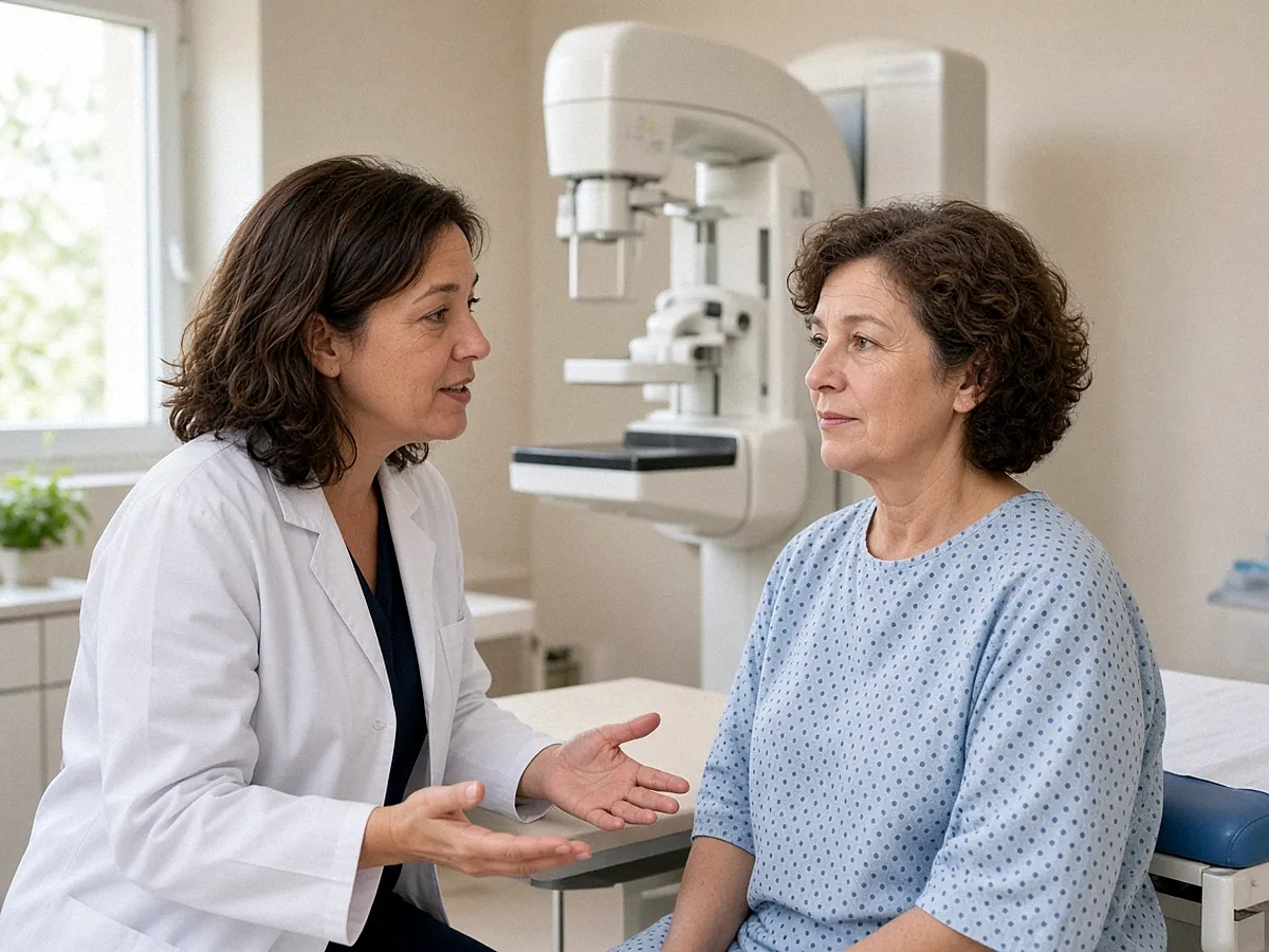
A repeat biopsy
A repeat biopsy is performed by the radiologist, under local anaesthetic. It most often uses a larger-gauge needle, or a vacuum-assisted biopsy, which draws in larger fragments and allows more tissue to be sampled:
- under stereotactic guidance, guided by mammography, for a cluster of microcalcifications or a distortion;
- under ultrasound, for a mass visible on ultrasound;
- under MRI, when the abnormality is visible only on MRI.
The time to the result depends on the laboratory. If this new result clearly explains the image, it may avoid an operation.
A reassuring biopsy is not always enough when the image remains suspicious: it is matching the two that establishes the diagnosis.
Surgical biopsy
Surgical biopsy, or diagnostic excision, consists of removing the whole suspicious area in order to analyse it. It is proposed when the discordance is clear-cut, when a repeat biopsy would not provide certainty, or when it is technically difficult.
The pathologist then analyses the whole lesion, and not just a few fragments: the diagnosis becomes final.
The decision at the multidisciplinary team meeting
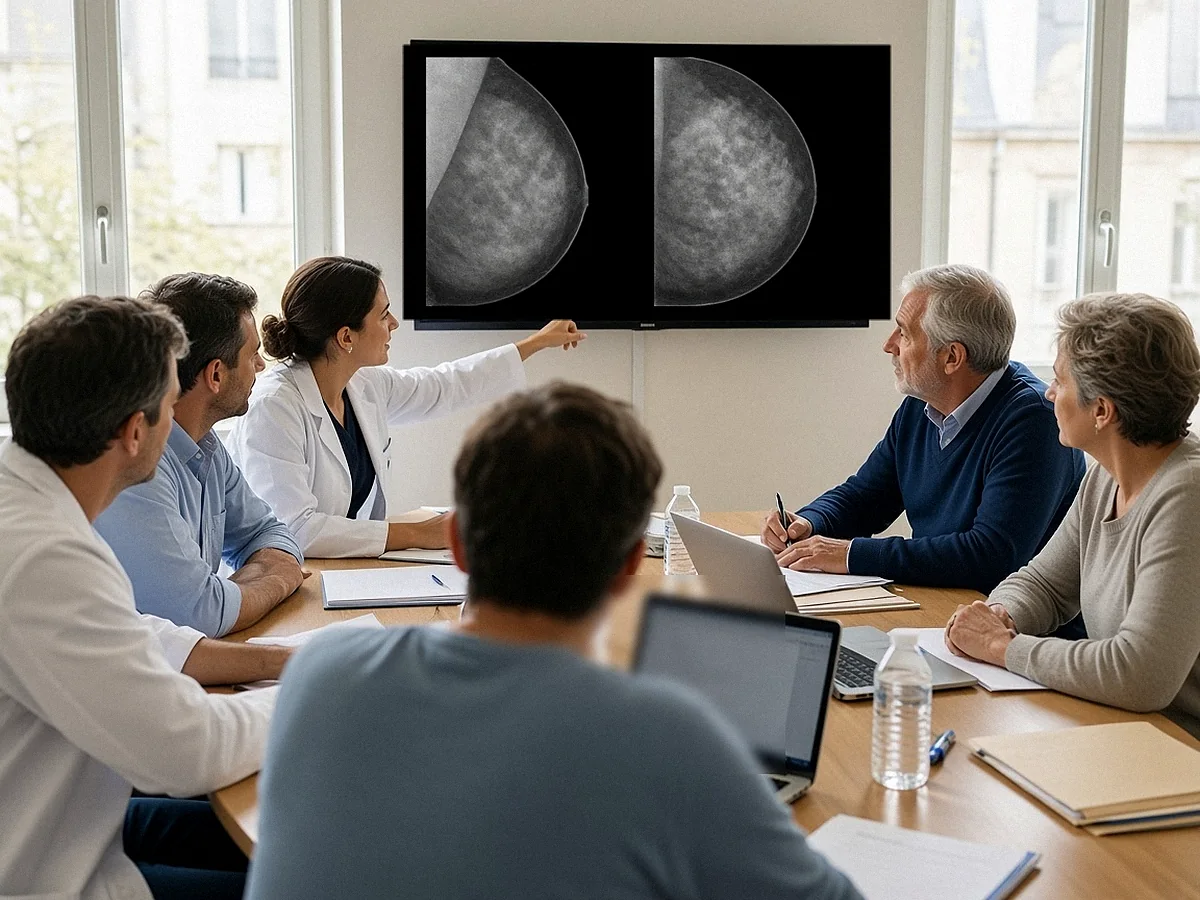
The choice between surveillance, a repeat biopsy and surgery is discussed at the multidisciplinary team meeting. This meeting brings together, in particular, the radiologist, the pathologist and the surgeon.
Each brings their own reading of the file: the radiologist presents the images and how the sampling went, the pathologist describes what was seen under the microscope, the surgeon assesses what an operation would add and how it would be performed. The opinion given is then explained to you in consultation, and the decision is made with you.
Dr Zeitoun reviews the imaging and the reports, examines you and explains why a check is being proposed, and in what form.
If a repeat biopsy is indicated, it is performed by the radiologist. Appointment on Doctolib, online consultation or request a callback from the secretariat.
B3 lesions: a neighbouring situation
A B3 lesion, described as of uncertain malignant potential, is not a cancer. But it may be associated with a nearby cancer that the biopsy did not sample, and some increase the risk of breast cancer later on.
This group includes in particular atypical ductal hyperplasia, lobular neoplasia, papillary lesions such as papilloma, radial scar and certain fibroepithelial lesions, including phyllodes tumour.
After a core needle biopsy showing a B3 lesion, excision is most often proposed. According to the 2023 international consensus and the 2024 European guidelines:
- for atypical ductal hyperplasia and phyllodes tumour, surgery remains preferred, because of a higher risk of finding a cancer in the specimen;
- for the other B3 lesions, either a vacuum-assisted excision, performed by the radiologist, or surgery is proposed, depending on your wishes, your age, the size of the lesion and your personal and family history;
- when the lesion has been completely removed, by vacuum-assisted biopsy or by surgery, and it is certain that everything has been removed, imaging surveillance is essential for several years.
These options are, here too, discussed at the multidisciplinary team meeting. See also benign breast surgery.
The operation: a diagnostic procedure
A surgical biopsy for discordance is a diagnostic procedure. It does not presume a cancer, and it is not a mastectomy: only the area in question is removed, with a small margin of tissue around it.
Preoperative localisation
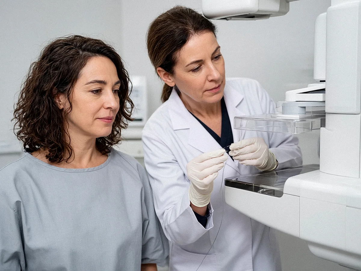
The lesion often cannot be felt. On the day of the operation or a few days before, the radiologist places a marker in the area to be removed, under ultrasound or mammography. This preoperative localisation is performed under local anaesthetic.
It may be a hookwire, a fine wire whose tip is anchored in the lesion, or a magnetic clip, depending on the case. The small metal clip placed during the biopsy often serves as the target.
In the operating theatre
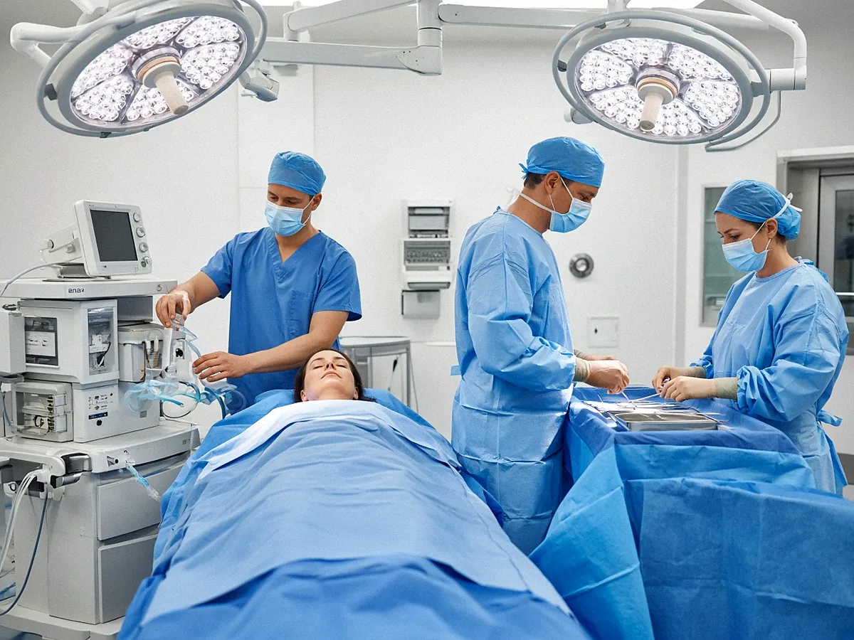
The surgeon removes the localised area, placing the incision as discreetly as possible, for example around the areola when the position of the lesion allows. The operation is performed under general anaesthetic, most often as day surgery, with discharge the same day.
During the operation, the removed specimen is X-rayed. This X-ray checks that the marker, the clip and, where applicable, the cluster of microcalcifications are indeed in the specimen, before it goes to the laboratory.
Dr Zeitoun practises in Sector 2 (non-OPTAM) and charges fees above the French Social Security rate for all consultations and procedures. Most international patients are not covered by French Social Security. A detailed quote is given during the consultation, before any procedure. If you are covered, the Assurance Maladie reimburses on the basis of the Social Security rate, and your health insurance may cover all or part of the additional fees, depending on your policy.
After the operation
Recovery is usually straightforward: some discomfort or moderate pain, relieved by standard painkillers, and sometimes a bruise that fades. See preparing for your operation and recovery after surgery.
The whole surgical specimen is analysed. The result is reviewed in consultation and, if needed, at the multidisciplinary team meeting.
What the result of the operation may show
Analysis of the surgical specimen most often shows a benign lesion. It may also show an at-risk lesion or a cancer.
A benign lesion
This is the most common case. The lesion is removed, the diagnosis is final, and follow-up resumes according to your age and your risk factors.
An at-risk lesion
Atypical ductal hyperplasia and lobular neoplasia are not cancers. They increase the risk of breast cancer later on. Appropriate surveillance is then proposed, and a cancer genetics consultation may be discussed depending on your family history.
A cancer
If the analysis shows a breast cancer, in situ or invasive, the next steps are discussed at the multidisciplinary team meeting. The surgical biopsy has often already removed the lesion; it is therefore a lumpectomy.
A second operation may be needed, to widen the safety margins if they are too close to the lesion, or to look for the sentinel lymph node in the case of an invasive cancer. Systemic treatment and radiotherapy are then discussed according to the characteristics of the tumour.
Follow-up after a biopsy or an operation
When a biopsy result is benign and concordant, no operation is generally needed. Depending on the nature of the lesion, the radiologist may propose follow-up imaging at 4-6 months before considering a return to routine follow-up.
After a surgical biopsy showing a benign lesion, follow-up resumes according to your age and your risk factors. After an at-risk lesion, such as atypical ductal hyperplasia, closer surveillance of both breasts is proposed.
Report without delay any new lump, any change in the skin or the nipple, or any bloody discharge.
What to expect from the consultation
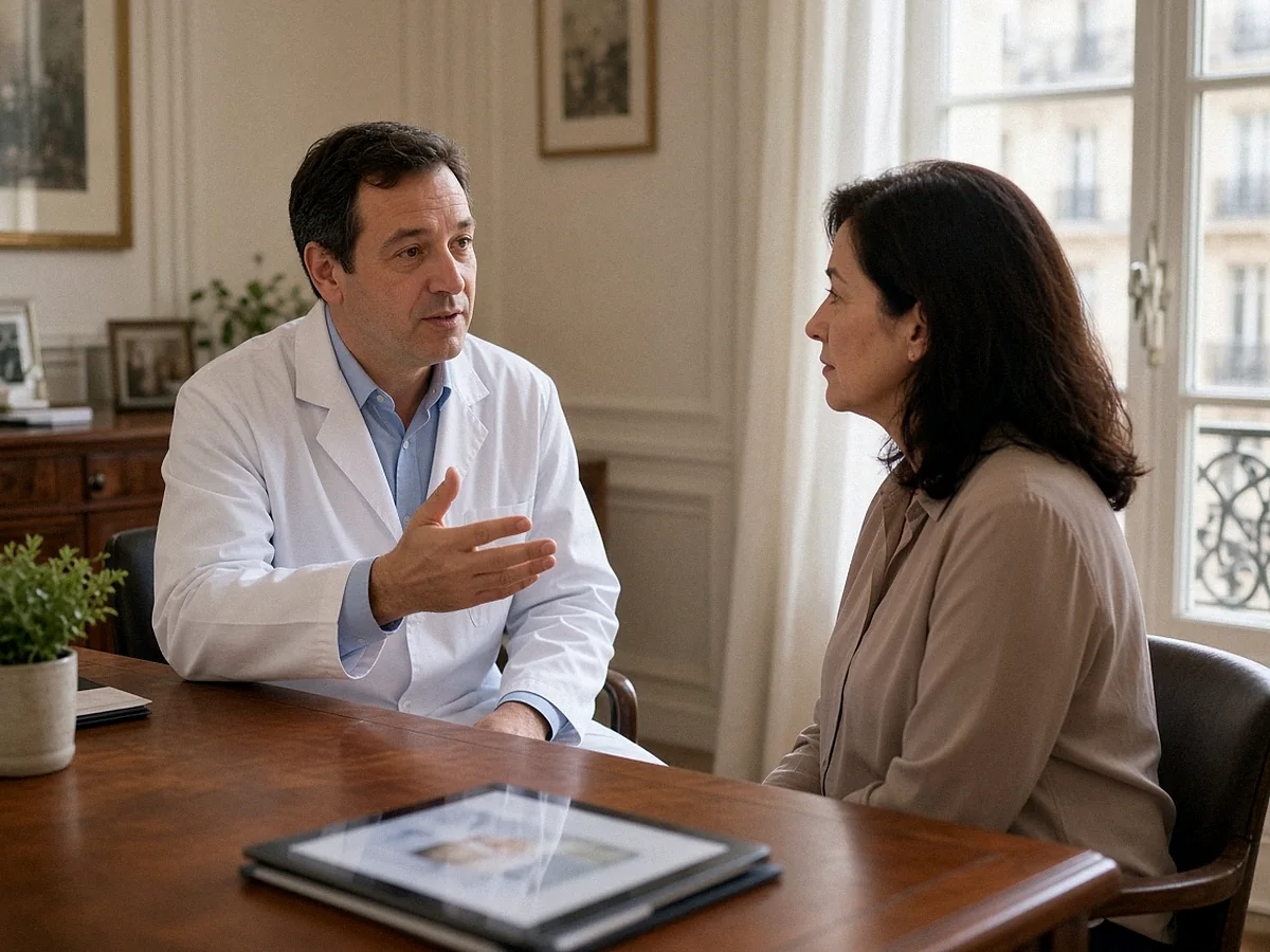
The consultation is used to review the whole file: the images, the biopsy report and the radiologist’s conclusion on concordance. Dr Zeitoun examines you, explains why a check is needed and compares the options with you, repeat biopsy or surgery.
Bring the films and CDs, the imaging and biopsy reports, and previous examinations if you have them. See Dr Zeitoun’s background, preparing your first consultation, second opinion and choosing a breast surgeon in Paris.
Dr Zeitoun sees patients at 241 rue du Faubourg Saint-Honoré (Paris 8th) and at Clinique Hartmann (Neuilly-sur-Seine).
Do you still have questions about your biopsy result or about a proposed operation?
Dr Zeitoun can go over the images, the report and the options with you. Online consultation, appointment on Doctolib, or request a callback from the secretariat.
Frequently asked questions
My biopsy is benign: why talk about surgery?
Because the result does not explain the image. When the image is highly suspicious or the target was not reached, a benign result is not enough to rule out a lesion. A repeat biopsy or an excision makes it possible to obtain a certain diagnosis.
Does discordance mean that I have cancer?
No. In most cases, the lesion turns out to be benign. But a cancer is found in a significant proportion of cases, which justifies completing the diagnosis.
Who decides that there is discordance?
The radiologist who performed the biopsy compares the pathologist’s result with the images. The file is then discussed at the multidisciplinary team meeting.
Can the biopsy be repeated rather than operating?
Often, yes. A vacuum-assisted biopsy under stereotactic or ultrasound guidance, performed by the radiologist, may be enough when its result clearly explains the image. Surgery is proposed when the discordance is clear-cut or when a repeat biopsy would not provide certainty. See breast biopsy.
How does the surgeon find a lesion that cannot be felt?
Thanks to a preoperative localisation placed by the radiologist, a hookwire or a magnetic clip. The specimen is X-rayed during the operation to check that the area has indeed been removed.
Is a B3 lesion a cancer?
No. It is a lesion of uncertain malignant potential. Excision is most often proposed, by surgery or by vacuum-assisted biopsy depending on the type of lesion. See atypical ductal hyperplasia.
When is follow-up imaging done after a benign biopsy?
When the result is benign and concordant, the radiologist may propose, depending on the nature of the lesion, follow-up imaging at 4-6 months before considering a return to routine follow-up.
Is the operation performed under general anaesthetic?
Yes. The surgical biopsy is performed under general anaesthetic, most often as day surgery, with discharge the same day. See recovery after surgery.
If the operation shows a cancer, is another operation needed?
Not always. The surgical biopsy has often already removed the lesion. A second operation may be needed to widen the safety margins or to look for the sentinel lymph node in the case of an invasive cancer.
What are Dr Zeitoun’s fees?
Dr Zeitoun practises in Sector 2 (non-OPTAM) and charges fees above the French Social Security rate for all consultations and procedures. Most international patients are not covered by French Social Security. A detailed quote is given during the consultation, before any procedure. If you are covered, the Assurance Maladie reimburses on the basis of the Social Security rate, and your health insurance may cover all or part of the additional fees, depending on your policy.
Can I ask for a second opinion?
Yes, on the images as well as on the biopsy report. See second opinion for breast cancer.
Sources
- SENORIF 2025-2026 guidelines — Breast cancers and breast diseases: diagnostic and therapeutic approaches. Public page of the guidelines
- Yincharoen P., Sharma C.P., Tawanwongsri W. — Radiologic–Pathologic Discordance After Image-Guided Breast Biopsy: A Systematic Review of Prevalence and Outcomes. Medical Sciences, 2026, 14(2), 229 (doi: 10.3390/medsci14020229). Twenty-three studies; discordant benign result in 1.2 to 5.3% of all biopsies; cancer found in about 20 to 40% of cases in representative series after re-sampling or excision.
- Flegg C. et al. — CAR Practice Guidelines on Breast Imaging and Interventions: Breast Intervention and Biopsy Procedures. Canadian Association of Radiologists Journal, 2026 (doi: 10.1177/08465371261451254). Concordance determined by the radiologist who performed the biopsy, discussion with the pathologist; X-ray of samples for microcalcifications; discordant benign result: repeat image-guided biopsy or surgical excision.
- Elfgen C. et al. — Third International Consensus Conference on lesions of uncertain malignant potential in the breast (B3 lesions). Virchows Archiv, 2023 (doi: 10.1007/s00428-023-03566-x). Surgical excision recommended for atypical ductal hyperplasia and phyllodes tumour; vacuum-assisted excision a possible alternative for the other B3 lesions; surveillance possible after complete removal by vacuum-assisted biopsy.
- Rubio I.T. et al. — European guidelines for the diagnosis, treatment and follow-up of breast lesions with uncertain malignant potential (B3 lesions) developed jointly by EUSOMA, EUSOBI, ESP (BWG) and ESSO. European Journal of Surgical Oncology, 2024, 50(1), 107292 (doi: 10.1016/j.ejso.2023.107292; PMID: 38061151). Multidisciplinary discussion to ensure radiologic-pathologic concordance; choice between vacuum-assisted excision and surgery according to atypia, lesion size, volume sampled and the patient’s preferences.
- The Royal College of Pathologists — Guidelines for non-operative diagnostic procedures and reporting in breast cancer screening (G150), 2021. Categories B1 to B5 for breast biopsies; after a B3 or B4 result, diagnostic excision or repeat biopsy, without treatment surgery on this result alone.