Home
Breast & breast cancer
Breast cancer Benign breast lesions Prophylactic mastectomy
Breast reconstruction
All techniques Implant reconstruction Latissimus dorsi flap DIEP flap (abdomen) Gracilis flap (thigh) Fat grafting (lipofilling) Flat closure Intimate surgery
Benign gynaecological surgery
Uterus Ovaries and tubes Cervix Vulva and vagina
Gynaecological cancers
Cervical cancer Ovarian cancer Uterine cancer Vulvar cancer Borderline tumours Articles About
Request a callback Book on Doctolib →

The words “radial scar” appear on the report from your biopsy. Despite this name, it is not an old scar: the term can appear in a woman who has never had breast surgery or a blow to the breast. It is a name given under the microscope, which describes the appearance of a small area of the gland.

The radiologist has performed the imaging and taken the sample. Dr Zeitoun, the surgeon, comes in next: he goes through your records with you and explains what follows. The explanations below are based on the SENORIF 2025-2026 reference, the reference document of the teams that care for breast diseases in the Île-de-France region.

Does your report mention a radial scar? Dr Zeitoun goes through the images and the results with you, then explains what is offered.

Request a callbackBook on Doctolib →
The microscope

A name given under the microscope, not an old scar

A pathologist at the microscope in a laboratory.

Under the microscope, the pathologist, the doctor who examines the sampled tissue, sees breast ducts converging towards a firm centre. It is this spoke-like appearance that gave the word “radial”.

The word “scar” comes from the appearance of this centre, which looks like scar tissue. It does not mean that the breast has been injured or operated on. A true scar, left by an earlier operation, is something else: tell the radiologist about any past operation on this breast.

A radial scar is a benign lesion, and not a cancer.

Why it is classed B3

The report from the pathologist places each sample in a category, from B1 to B5: B1 is normal tissue, B2 a benign lesion, B3 a benign lesion that needs further assessment, B4 a suspicious lesion and B5 a cancer.

This B3 has nothing to do with the ACR 3 category of the BI-RADS classification, which you may have read on a mammogram report. ACR 3 is given by the radiologist, from the image: it means an image that is probably benign, which is usually monitored with follow-up imaging rather than sampled. B3 is given by the pathologist, from the tissue removed at the biopsy: it means a benign at-risk lesion. The two 3s do not measure the same thing.

The radial scar is classed B3. It is called an at-risk lesion, or a lesion of “uncertain malignant potential”. This phrase does not mean that the area is in the process of becoming malignant. It is a reminder that the needle has analysed only part of the area: abnormal cells, or a cancer, may lie very close by without having been sampled. Other lesions are classed B3, such as a papilloma.

The report also states whether atypia is present. Atypia is a cell whose appearance differs from that of a normal cell, without being a cancer cell. Atypia beside the radial scar, for example atypical ductal hyperplasia, is taken into account in the decision.

Radial scar, complex sclerosing lesion, Aschoff nodule

These three names refer to the same family of lesions. European recommendations speak of a radial scar when the area is small, most often less than one centimetre, and of a complex sclerosing lesion when it is larger and less regular. “Aschoff nodule”, or Aschoff proliferative centre, is an older name, still used by some doctors. Care is discussed in the same way for all three.

Where it comes from

Where does a radial scar come from?

The precise cause is not known. It is not linked to a blow or to an earlier operation: the word “scar” describes only how it looks under the microscope.

It is usually discovered on a mammogram, often done as part of screening. It may also be found by chance, during a biopsy done because of another image.

The images

What you may feel, and what the images show

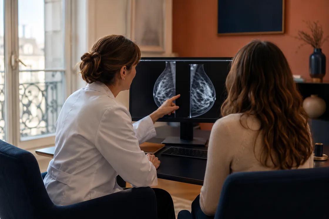
A radiologist in front of mammogram screens, in a reading room.

Most often, a radial scar causes no symptoms: no lump under the fingers, no discharge from the nipple, no change in the skin. It is found on imaging.

On the mammogram, it often appears as an architectural distortion, that is a small area where the lines of the gland come together towards one point, or as a spiculated image, star-shaped. It may also come with microcalcifications, tiny deposits of calcium visible on the images.

The radiologist often completes the work-up with an ultrasound, and sometimes with an MRI. The image is then classed with the BI-RADS system, the current name of the ACR classification, established by the American College of Radiology, which indicates whether an image is benign, to be monitored, or to be sampled.

What the radiologist sees, and what the microscope sees

Architectural distortion and the star-shaped image are appearances that the radiologist sees on the images. The radial scar is what the pathologist sees under the microscope, on the sampled tissue. The same distortion may correspond to a radial scar, to another benign lesion or to a cancer. The radiologist offers a biopsy in order to tell the difference.

The report

From the image to the report: the work of the radiologist

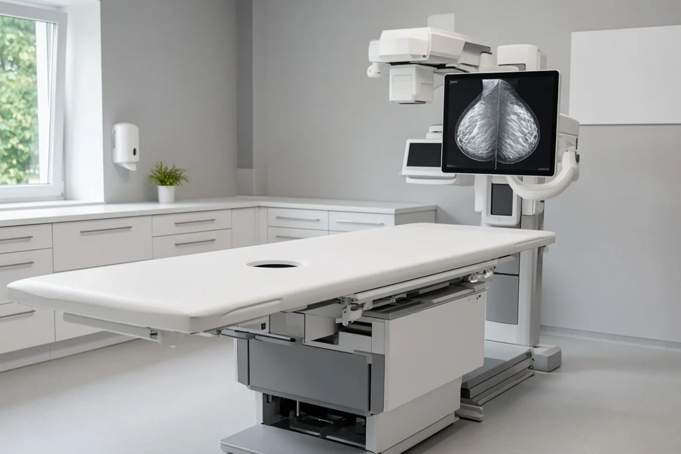
A vacuum-assisted biopsy room with a stereotactic table, and no patient.

The biopsy is a procedure performed by the radiologist, under local anaesthetic.

The radiologist chooses between two procedures. With a core needle biopsy, a fine needle is introduced several times and a core is collected at each pass, that is a thin cylinder of glandular tissue; ultrasound is most often the guide. With a vacuum-assisted biopsy, the needle is larger and draws the tissue in, which makes it possible to collect a larger volume without withdrawing it each time.

The radiologist finishes by placing a clip, a small piece of metal that marks the place that was sampled. It stays in place without discomfort and makes it possible to find the area again if an operation is decided. The report usually arrives within a few days. The reasons for this waiting time are set out in the article on how long a biopsy result takes.

Concordance: do the image and the microscope say the same thing?

When the report arrives, the radiologist checks that what the microscope says matches what was seen on the image. This is called concordance. If the two agree, for example a radial scar for a star-shaped image, this result can be relied on. If they do not agree, the case is discussed at an MDT meeting, the multidisciplinary team meeting at which several breast specialists review each case together. A new sample or removal of the area may then be offered.

The operation

Why an operation is most often offered

A small metal localisation clip resting on a drape.

A radial scar is benign. But it is one of the at-risk lesions, and each case is discussed at an MDT meeting. Most often, surgical excision is offered, that is removal of the area in the operating theatre, so that the whole of it can be analysed, and so that no cancer is missed immediately beside what the needle sampled. This offer is discussed case by case.

The MDT meeting, then the decision with Dr Zeitoun

At the MDT meeting, radiologists, pathologists, surgeons and oncologists (doctors who specialise in the treatment of cancer) examine your images and your reports together. After this meeting, Dr Zeitoun explains the proposal to you and decides the next step with you.

Three possibilities exist, depending on the case:

  • surgical excision, the option most often offered;
  • vacuum-assisted excision, performed by the radiologist, which removes the whole image with the needle, without going to the operating theatre;
  • surveillance with imaging.

The choice is made case by case, according to the appearance of the image, whether or not atypia is present, your age, your medical history and your wishes.

The operation

The area is usually not palpable, which means it cannot be felt by touching the breast. Before the operation, the radiologist therefore places a marker in contact with the clip: this is preoperative localisation, the techniques for which are presented on that page. Dr Zeitoun then removes the marked area. The procedure corresponds to a lumpectomy, the removal of a small part of the breast; how it is performed and what follows are set out on that page. The operation takes place at Clinique Hartmann, in Neuilly-sur-Seine.

To understand the decision taken at the MDT meeting or to prepare for the operation, Dr Zeitoun sees you with your records.

Request a callbackBook on Doctolib →
After the operation

What you should expect after the operation

The whole of the removed area is analysed by the pathologist. Dr Zeitoun explains this result to you at a consultation after the operation.

Most often, the analysis confirms a radial scar, with or without atypia. In that case, there is usually no reason to operate again, even if the lesion touches the margins, that is the edges of the removed tissue.

The analysis may sometimes show a cancer. The case is then discussed again at the MDT meeting and Dr Zeitoun presents the appropriate treatment to you. If the margins of the removed tissue are not healthy, it may be necessary to operate a second time.

Breast surveillance afterwards

Surveillance by ultrasound and/or by mammogram is then planned for ten years, at intervals agreed with Dr Zeitoun.

The risks of the operation

Removal of a small area of the breast is a common operation. Possible after-effects, such as a haematoma (a small pocket of blood under the scar), are described on the lumpectomy page and gone through with you before the operation.

The appointment

What you should expect from your surgical appointment

Mammogram images, reports and an imaging disc laid on a desk.

The appointment takes place after the biopsy. Dr Zeitoun reviews the images, the report from the radiologist and the report from the pathologist, and presents the proposal made after the MDT meeting. If an operation is chosen, he explains the localisation, where the scar will sit on the skin, and when the analysis of the removed area will be available.

Please bring your complete imaging records: the images and the disc, the reports from the radiologist, the biopsy report, the examinations from earlier years so they can be compared, and your current prescriptions.

You have already received a proposal and would like another view: Dr Zeitoun can give a second opinion on your case.

Consultations at the practice, 241 rue du Faubourg Saint-Honoré (Paris 8th), and at Clinique Hartmann (Neuilly-sur-Seine). Surgery takes place at Clinique Hartmann.

Sector 2, not opted into OPTAM, with possible fee supplements. The amount is explained during the consultation and written on a quote before the procedure.

Questions

The questions most often asked about a radial scar

I have not had breast surgery. Why is the word scar used?
▾

It is a name read on the biopsy report, which only describes the appearance under the microscope: ducts that converge towards a firm centre, similar to scar tissue. It implies neither an operation, nor a blow, nor an injury.

Is a radial scar a cancer?
▾

No. It is a benign lesion classed B3, among the at-risk lesions. The operation, when it is offered, serves to check that there is no cancer beside the area sampled by the needle.

Is the operation compulsory?
▾

Not necessarily. Most often, surgical excision is offered. Depending on the case, the MDT meeting may also choose a vacuum-assisted excision performed by the radiologist, or surveillance. Dr Zeitoun decides with you, case by case.

Does a biopsy have to be repeated before the operation?
▾

Most often, no: the operation is decided after the MDT meeting, on the basis of the first biopsy. A new sample may be discussed if the result does not match the image.

What happens if the edges of the removed tissue are not healthy?
▾

If the analysis shows a radial scar, with or without atypia, there is usually no reason to operate again. If it shows a cancer, the case is discussed again at the MDT meeting, and a second operation may be needed in order to obtain healthy edges.

What surveillance follows a radial scar?
▾

Surveillance by ultrasound and/or by mammogram for ten years, at intervals agreed with Dr Zeitoun.

Sources

  • SENORIF reference. Cancers et pathologies du sein: attitudes diagnostiques et thérapeutiques, protocoles de traitement 2025-2026 (updated July 2026), chapter “Lésions de potentiel malin incertain (B3)”.
  • French National College of Gynaecologists and Obstetricians (CNGOF). Lavoué V et al. Tumeurs bénignes du sein: recommandations pour la pratique clinique, short text. J Gynecol Obstet Biol Reprod (Paris). 2015;44(10):1049-1064.
  • Rubio IT et al. European guidelines for the diagnosis, treatment and follow-up of breast lesions with uncertain malignant potential (B3 lesions) developed jointly by EUSOMA, EUSOBI, ESP (BWG) and ESSO. Eur J Surg Oncol. 2024;50(1):107292.
  • Elfgen C et al. Third International Consensus Conference on lesions of uncertain malignant potential in the breast (B3 lesions). Virchows Arch. 2023;483(1):5-20.
  • Pinder SE et al. NHS Breast Screening multidisciplinary working group guidelines for the diagnosis and management of breast lesions of uncertain malignant potential on core biopsy (B3 lesions). Clin Radiol. 2018;73(8):682-692.

Further reading

Dr Zeitoun sees you to go over your report and organise what follows, at the practice or at Clinique Hartmann.

Request a callbackBook on Doctolib →
Former resident at the Centre François Baclesse (Caen) and the Institut Curie; former specialist practitioner in the French comprehensive cancer centres at Gustave Roussy.
Page reviewed by Dr Jérémie Zeitoun on 7 October 2026, at 6:38 pm.
FREN
ONLINE CONSULTATIONVideo consultation · by appointment