The mammogram report mentions microcalcifications. The word describes a focus seen on the image — not a diagnosis. On its own it does not say whether surveillance, a biopsy, or an operation is the right next step. Here is how to read that line, and when a breast surgeon joins the pathway.
Dr Jérémie Zeitoun sees patients in consultation in Paris and/or in Neuilly (Clinique Hartmann), with the mammogram and, if it already exists, the biopsy report. When an operation is decided, it takes place at Hartmann.
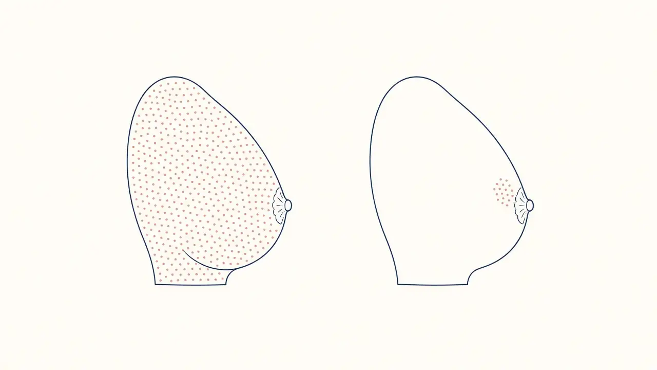
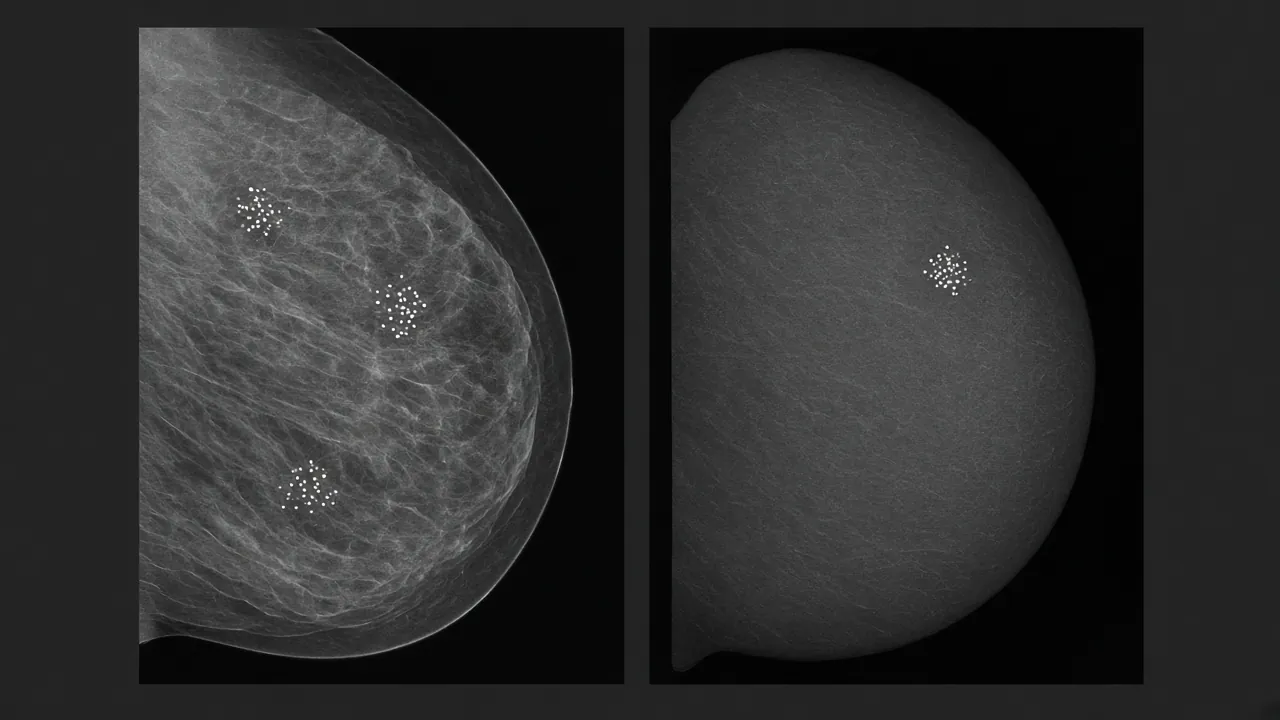
A mammogram can describe microcalcifications when no lump has been felt. They are very small points of calcium in breast tissue, visible on the film. They are not, by themselves, a diagnosis of breast cancer.
Many of them go with benign change: ageing of the ducts, an old scar, a blood vessel, the skin. Other patterns, especially a small irregular cluster in one area, lead the radiologist to recommend a sample. Only the tissue analysis says what is actually there.
This page stays with the report and the decision that follows: surveillance, a biopsy, or an operation. It does not repeat how the sample is taken, the full BI-RADS scale, or the treatment of ductal carcinoma in situ once that diagnosis is written. Those three subjects already have their own pages.

The breast is made of ducts, lobules and supporting tissue. Calcium can settle there in amounts too small to form a lump you can feel. Mammography is the test that shows them best. Ultrasound, which is very useful for a mass, often does not show isolated microcalcifications. A “normal” ultrasound therefore does not cancel the mammogram report.
Their presence does not state their nature. The same white speck can sit beside benign change, atypia — cells a little different from normal, without being a cancer — a carcinoma still confined to the ducts, or, less often, a cancer that has crossed the duct wall. The sample, and sometimes the operation, is what settles it. The radiology report only points to the next step.
Comparison with earlier mammograms weighs as much as today’s film. A focus that has been stable for years does not carry the same meaning as a new focus. If the breasts are dense, reading the film is harder. Microcalcifications can still be the finding mammography is able to show.
They are usually not felt. They do not replace the pathology report. A line that mentions them calls for a reading, not an immediate conclusion.
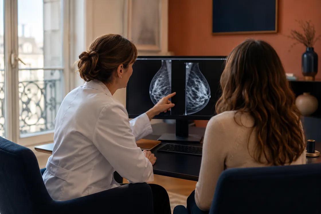
The written report is a summary. The decision rests on the films themselves, on disc or online, and on earlier examinations. Preparing the first consultation lists what is useful to have.

The report describes two separate things: where the microcalcifications sit, and whether the appearance is reassuring or not. Distribution can be said in ordinary language. Shape sometimes uses radiology words. Neither one replaces the final category on the report, which states the level of concern and the step recommended. That category is explained on the page mammogram BI-RADS.
Some images are recognised as benign from their appearance alone: calcium in the skin, calcium that follows a blood vessel, coarse “popcorn” calcifications in an old fibroadenoma, rod-shaped calcifications linked to ageing of the ducts, or known traces of earlier surgery or radiotherapy. When they are typical, they do not lead to a biopsy. When a doubt remains, the radiologist says so, and recommends a sample rather than an open-ended wait.

At the end of the report there is a category, called BI-RADS, sometimes still written as ACR. It does not name a disease. It states the radiologist’s level of concern and the step recommended. The full scale, from an incomplete assessment to a cancer already proved, is on the page what happens after a BI-RADS 4 or 5 mammogram. For microcalcifications, three situations come up again and again.
The appearance is benign, or judged probably benign. You return to screening mammography, or to a check whose date the report sets, often a few months later. Nothing is biopsied while the image stays in that frame.
The appearance is suspicious: a new cluster, irregular shapes, the course of a duct. A sample is proposed. A breast radiologist performs it. It is not an operation.
It is not decided from the mammogram alone. It is discussed after the biopsy result, when that result shows atypia, carcinoma in situ, or a cancer.
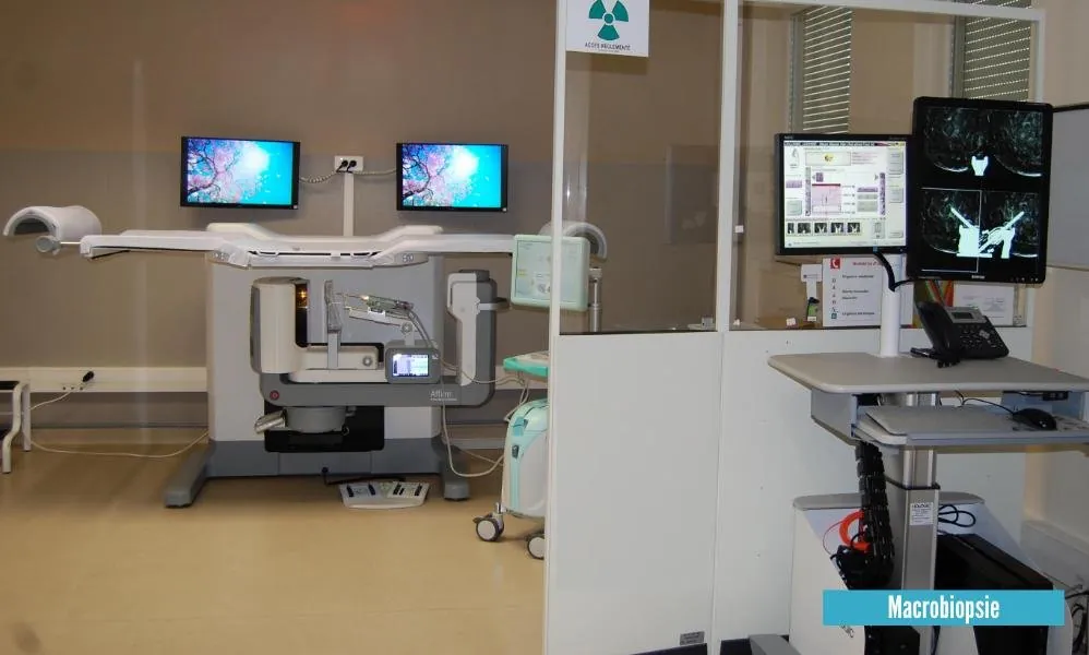
If ultrasound shows no target, the focus cannot be aimed at “blind”. The usual procedure is then a stereotactic vacuum-assisted biopsy: the breast is compressed as for a mammogram; the radiologist locates the focus on the films, then samples, under local anaesthetic, several tissue fragments with the aspiration needle. They are called “cores”: small cylinders of breast tissue for pathology. These cores are X-rayed straight away, to confirm that the targeted focus is inside them. A small metal clip is often left in place, so the area can be found again later.
How the procedure feels and how to read the result (the B classification) are on the page breast biopsy. The radiologist performs this sampling himself. Dr Zeitoun then reads the file with you to say whether surveillance or an operation is required.
Two readings matter as much as the word “benign”: concordance or discordance between the image and the histology. Concordance: the focus was in the cores, and the result matches what the mammogram showed. Discordance: the image was highly suspicious, or the cores do not contain the targeted microcalcifications — another sample is then discussed before any conclusion.
Mammogram alone, biopsy already done, or an operation already proposed elsewhere: the consultation (Paris and/or Neuilly — Hartmann) is there to say which step is the right one; surgery, if retained, takes place at Hartmann.

The result usually arrives within a few days to two weeks. It describes the tissue sampled, not “the microcalcifications” as if they were a single disease. Three outcomes guide the surgical consultation. The detail of the B categories is on the page breast biopsy. Here, only the consequence for a woman whose finding was a cluster of microcalcifications.
The tissue is benign alone, and radio-histological concordance is assured (the focus was in the cores). There is no operation. Follow-up at 6 months for microcalcifications (at 4 months if it was a mass). The foci can remain visible: a benign result does not erase them.
The most frequent example on a cluster of microcalcifications is atypical ductal hyperplasia: duct cells a little different from normal. It is not a cancer. The needle has seen only part of the area. Removing the marked site is often proposed, because examination of the whole specimen can find, beside the atypia, a carcinoma in situ or, less often, an invasive cancer. It can also confirm atypia alone.
Ductal carcinoma in situ is treated with surgery: breast conservation when the cluster is limited, mastectomy when it occupies too much of the breast. An invasive cancer opens the breast cancer pathway, with a multidisciplinary meeting. Both diagnoses have their own page. This one does not repeat their treatment.
Other findings — a radial scar, a papilloma, flat epithelial atypia — are discussed in the same way: does the sample represent the area, and should the rest of it be removed in order to be sure? The answer is given on the images and the report, not by a single rule.
The consultation is in Paris and/or in Neuilly (Clinique Hartmann). Surgery takes place at Hartmann. A mammogram done in another city, in France or from abroad, is a valid starting point: examinations are not repeated if they are complete and readable. A video consultation allows a first contact to arrange sending the file. The decision to operate is confirmed after a clinical examination, not from a message alone.
When the diagnosis is carcinoma in situ or a cancer, the plan goes through a multidisciplinary meeting. The operation removes the marked area. It does not “treat” microcalcifications as dust that could be made to vanish: it removes the tissue in which they sit, so that tissue can be examined in full. The specimen report, given at a consultation, says whether the margins are clear and whether the nature confirmed is the one the biopsy announced.
Write to the officeThe questions that come up when the word “microcalcifications” appears on a report. If yours is not here, it belongs in the consultation — or with Sophie, at the bottom right of the screen.
No. They are foci of microcalcifications. Most of them go with benign tissue. Some patterns justify a biopsy, and it is that analysis — not the word on the report — that says whether there is atypia, carcinoma in situ, or a cancer.
As a rule, no. They do not form a lump. A mass you can feel is a different situation, looked at with ultrasound and, if needed, a biopsy aimed at that mass. The two can exist together; the report says so when they do.
Scattered microcalcifications, often in both breasts, with a regular appearance, are usually kept under surveillance. A cluster in a small area, especially if it is new or irregular, or if it follows a duct, generally leads to a sample. The BI-RADS category on the report states that recommendation.
No, if the mammogram describes a cluster that should be sampled. Ultrasound often does not show microcalcifications when there is no mass. That is why the biopsy is stereotactic, and not skipped because the ultrasound was reassuring. Dense breasts do not change this logic.
It means a biopsy is indicated. It does not make the diagnosis. Some of these samples come back benign, some show atypia or carcinoma in situ, some an invasive cancer. The page on BI-RADS 4 or 5 describes the weeks that follow that category, whether the image is a mass or calcifications.
Stereotactic vacuum-assisted biopsy is performed by a breast radiologist: he performs it himself, under local anaesthetic. Dr Zeitoun then reads the result with you and decides on any surgical next step. The procedure is described on the page breast biopsy.
Not always. Sometimes the result is benign but at risk (atypical ductal hyperplasia, papilloma, radial scar…): an operation may then be indicated. If the result is truly benign alone, and after radio-histological concordance is assured, follow-up is arranged: at 4 months for a mass, at 6 months for microcalcifications. Benign foci usually remain visible: the check compares them; it does not wait for them to fade.
Because the needle has examined only a fraction of the cluster. Removing the marked area allows the whole of it to be analysed. That examination sometimes finds carcinoma in situ or, less often, an invasive cancer right beside the atypia. It also often confirms that atypia was all there was. The operation answers that uncertainty. It is not proposed in order to “remove calcifications” as such.
It depends on how far the microcalcifications extend and on the size of the breast. A limited cluster lends itself to breast conservation, sometimes with oncoplastic surgery. Widespread disease leads to a discussion of mastectomy, with or without reconstruction. The detail of this diagnosis is on the page ductal carcinoma in situ.
Yes. Bring the images and the reports. The consultation is in Paris and/or in Neuilly (Hartmann); surgery takes place at Hartmann. The office explains how to send the file before you come. Dr Zeitoun’s training and practice are on the site.
No, if the films are recent, complete and obtainable. An examination is repeated only if a view is missing, if the images cannot be read, or if the radiologist has already asked for it. The useful list is on preparing the first consultation.
No. Benign tissue can go on carrying these foci of microcalcifications. They then serve as a point of comparison. The relief is about the nature of the tissue, not about the image fading. The next check is a mammogram, on the date given.
This page is separate from breast biopsy (the procedure and how to read the result), from the BI-RADS mammogram page (the weeks after category 4 or 5, whether the image is a mass or calcifications) and from ductal carcinoma in situ (treatment once that diagnosis is written).
Neighbouring pages, so the same subject is not read twice.
What the category changes in the weeks that follow, whether the image is a mass or calcifications.
The sampleHow a vacuum-assisted biopsy unfolds, and how the result is read.
If that diagnosis is madeWhat the diagnosis means, and how the operation is chosen.
The operationHow the choice is made when the area to remove is limited or extensive.
A different situationA mass you can feel is not read the same way as a cluster of microcalcifications.
Before you comeImages, reports, earlier examinations: what is useful to have to hand.
A report of microcalcifications, a biopsy already done, or an operation already proposed: Dr Jérémie Zeitoun sees patients in consultation in Paris and/or in Neuilly (Hartmann) and operates at Hartmann. Bring the mammogram and, if it exists, the pathology report.