Breast imaging · Breast surgery
Architectural distortion on a mammogram:
what happens next?
Further imaging, biopsy, reading the result and, when it is offered, the operation: the pathway step by step.
Further imaging, biopsy, reading the result and, when it is offered, the operation: the pathway step by step.
After the imaging and biopsy, performed by the radiologist, Dr Zeitoun sees you in consultation to explain the result and the next steps. Consultations at the practice, 241 rue du Faubourg Saint-Honoré (Paris 8th), and at Clinique Hartmann (Neuilly-sur-Seine). Surgery takes place at Clinique Hartmann.
This page is for people whose mammogram, ultrasound or MRI report mentions architectural distortion. The term is worrying, because it often leads to a biopsy. Yet it describes an image, not a diagnosis: several causes, benign or not, can produce it. The SENORIF guidelines, based on the French national recommendations, frame each step described here. Imaging and biopsy are performed by the radiologist; Dr Zeitoun then sees patients in consultation, usually with the biopsy result.

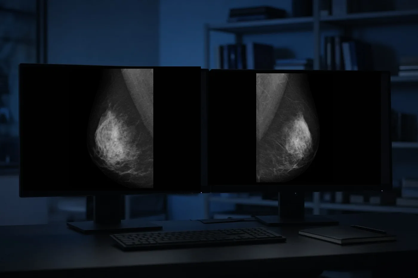
On a mammogram, the breast gland has a regular organisation: fine bands of tissue start from the nipple and spread out through the breast, a little like the ribs of a fan. Architectural distortion means that, in a small area, this organisation is pulled towards a point, like gathered fabric, without any visible lump.
What sets this image apart is that it is visible without a mass, meaning there is no growth at the point where the tissue is pulled. This is why nothing can usually be felt on examination.
In the ACR BI-RADS classification (American College of Radiology), an unexplained distortion, for example with no previous surgery at that site, is usually classified as ACR 4. This category means that a check under the microscope is recommended: it does not mean that cancer is present.
The same image can correspond to very different situations. A biopsy is the only way to tell them apart with certainty; the radiological appearance gives an indication, but is not enough on its own.
The share of each cause varies with age, medical history and the exact appearance of the image. The radiologist places your own case in context by looking at your images; the surgeon then reviews it, with the biopsy result.
Before taking a sample, the radiologist tries to see the area better, measure its extent and choose the simplest guidance for the biopsy.
Additional views are often taken first, with the mammography machine. A magnified view is centred on the area and shows it larger, so that its details can be seen better. A spot compression presses on that area alone with a small paddle, which separates overlapping tissue. If the image was only caused by normal tissue overlapping, it disappears on this view, which avoids an unnecessary biopsy.
Tomosynthesis, or three-dimensional mammography, is done with the same machine as the mammogram. Several images are taken at different angles while the tube moves in an arc above the breast; the computer then reconstructs them, which prevents tissue from overlapping. A distortion often shows up more clearly than on a standard view, and tomosynthesis can sometimes guide the biopsy.
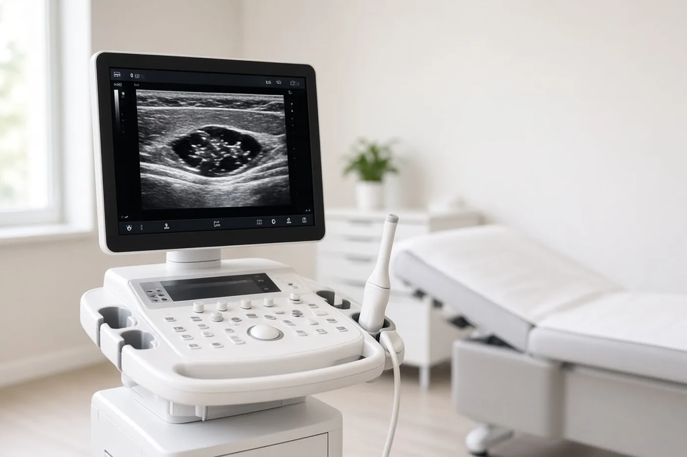
A targeted breast ultrasound looks for a match to the image, meaning that it tries to find on ultrasound the same abnormality as on the mammogram, in the same place in the breast: for example an ill-defined area, sometimes with a shadow behind it. If it finds it, the biopsy will usually be done under ultrasound, which is more comfortable.
A breast MRI is not routine. It is considered when the image is seen on only one view, that is, from only one of the two angles at which the mammogram is taken, or when its extent must be clarified before an operation. On MRI, axial, that is horizontal, slices compare both breasts at the same level.
At the end of this work-up, the report gives a final BI-RADS category. A category 4 or 5 leads to a biopsy.
A biopsy removes a few fragments of the area to analyse them under the microscope. It is performed by the radiologist, under local anaesthetic, as an outpatient, that is, without a night in hospital.
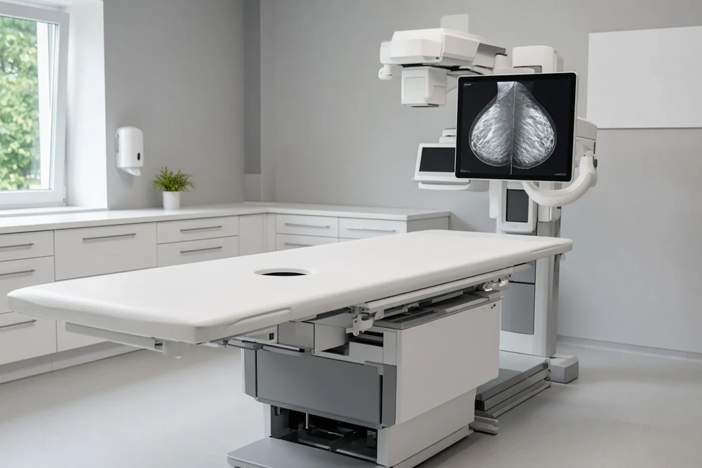
There are two techniques. A core needle biopsy uses a fine needle that takes several cores of tissue, meaning small cylinders of tissue the size of a pencil lead; it is most often guided by ultrasound. A vacuum-assisted biopsy uses a larger needle connected to suction, which removes more tissue in a single insertion; it is often guided by mammography or tomosynthesis when the image is not visible on ultrasound.
The choice depends on how visible the image is and on its size. For a distortion, a vacuum-assisted biopsy is often preferred, because it provides more material to analyse and makes the diagnosis more reliable. The procedure is described in detail on the page on breast biopsy.
At the end of the procedure, the radiologist leaves a clip in place, a tiny harmless metal marker. It marks the exact site of the sample and will serve as a guide if an operation is decided. A check mammogram confirms its position.
The result comes back after a few days; the page on how long biopsy results take explains why this time is needed and what can lengthen it.
The pathologist, the doctor who analyses tissue under the microscope, classifies the sample from B1 (normal tissue) to B5 (cancer). The decision never rests on this number alone: it takes into account whether the image and the result match.

Concordance is the central question. The radiologist checks that the result truly explains the image seen on the mammogram. A clear distortion with a result showing only normal tissue is called discordant: in this case, a repeat biopsy or an excision (removal of the whole area for analysis) is offered, even if the result is benign.
The page on pathology explains how this report is produced.
A B3, B4, B5 or discordant result is discussed at a surgical consultation, with your images and the biopsy report: after the multidisciplinary team meeting, Dr Zeitoun decides whether surgery is needed.
An operation is offered when the biopsy does not allow a sufficiently certain conclusion, or when it has shown cancer. In both cases, the procedure is targeted at the marked area.

A distortion cannot be felt under the fingers and cannot be seen with the naked eye during the operation: localisation is therefore essential. It shows the surgeon the exact area to remove, so that the right area is removed without taking more tissue than necessary. Its different forms are described on the pre-operative localisation page.
Two situations must be distinguished. For a B3 lesion or a discordance, this is a diagnostic excision: the area is removed in full so that it can be analysed completely and a certain diagnosis obtained. For cancer, this is a treatment: the lesion is removed with a margin of healthy tissue.
The course of the operation and recovery is explained on the lumpectomy page; localisation is covered on the pre-operative localisation page.
The surgical consultation takes place after the imaging and, usually, after the biopsy, both performed by the radiologist. It is the time to go through all your records with you, explain the recommended course of action and organise each step, without rushing.
The surgeon reviews the images, the imaging report and the biopsy result, checks concordance and explains why follow-up, a repeat biopsy or an operation is proposed. The surgeon also explains the type of localisation, the length of the operation, the planned scar and when the final result will be known.
Documents to bring: your mammogram, ultrasound and MRI images, with the CD if you have it; the reports; the biopsy result; your previous examinations, which help to judge any change; the list of your medication.
If you have already been given an opinion and would like to compare it, a second opinion is possible and is part of the usual pathway.
Consultations at the practice, 241 rue du Faubourg Saint-Honoré (Paris 8th), and at Clinique Hartmann (Neuilly-sur-Seine). Surgery takes place at Clinique Hartmann.
Sector 2, not opted into OPTAM, with possible fee supplements. The amount is explained during the consultation and written on a quote before the procedure.
No. A distortion can come from an old scar, a radial scar or sclerosing adenosis, which are benign. It can also reveal a cancer, which is why a biopsy is recommended when the image is not explained.
The distortion is seen on imaging without forming a mass, so it can rarely be felt. The absence of a lump does not allow a conclusion, and only the microscopic analysis of a sample gives a reliable answer.
It is a benign lesion in which a small fibrous centre draws in the neighbouring ducts, unrelated to any operation. It is a high-risk lesion: because the biopsy may miss a cancerous lesion lying next to it, surgery is most often needed. After the multidisciplinary team meeting, Dr Zeitoun decides whether to operate. The page on benign breast lesions describes it in detail.
“Without a mass” means that no lump is visible at the point towards which the tissue is pulled. This detail guides the radiologist, but does not replace a biopsy.
It depends on concordance. If the result truly explains the image, follow-up is usually enough. If it does not, or if it shows a B3 lesion such as a radial scar or atypia, Dr Zeitoun decides after the multidisciplinary team meeting whether surgery is needed; an excision is most often offered.
The course of the operation is explained on the lumpectomy page, and localisation on the pre-operative localisation page. The length of time off work depends on your job and on the procedure performed; it is agreed with you in consultation.
The distortion cannot be felt and is not visible to the naked eye during the operation. Localisation shows the surgeon the exact area to remove, so that the right area is removed without taking more tissue than necessary. Its different forms are described on the pre-operative localisation page.
The radiologist first tries to find it on ultrasound, known as a second-look ultrasound. If it remains invisible, an MRI-guided biopsy is performed. The page on breast MRI explains how it is done.
The biopsy result is usually known after a few days, as explained on the page on how long biopsy results take. When an operation is indicated, it is arranged in the following weeks, without haste but without unnecessary delay.
The consultation takes place after the imaging and biopsy, performed by the radiologist. Consultations at the practice, 241 rue du Faubourg Saint-Honoré (Paris 8th), and at Clinique Hartmann (Neuilly-sur-Seine). Surgery takes place at Clinique Hartmann. Sector 2, not opted into OPTAM, with possible fee supplements. The amount is explained during the consultation and written on a quote before the procedure.
Sources: SENORIF 2025-2026 guidelines (Île-de-France regional cancer network); Haute Autorité de santé (HAS, French National Authority for Health), breast cancer screening and diagnosis; Institut national du cancer (INCa, French National Cancer Institute), breast cancer diagnostic pathway; American College of Radiology, ACR BI-RADS Atlas, 5th edition.
After the biopsy, to go through the result with the surgeon or prepare for surgery: book on Doctolib, or request a callback from the secretariat.