
Sclerosing adenosis
Sclerosing adenosis:
understanding this biopsy result
A benign breast lesion, sclerosing adenosis is identified under the microscope, in the tissue removed during a biopsy.
A benign breast lesion, sclerosing adenosis is identified under the microscope, in the tissue removed during a biopsy.
It can be simple or complex, with or without atypia. Without atypia, it is classified B2: in the pathologist's categories (the pathologist is the doctor who analyses the tissue), B2 means that the lesion seen under the microscope is benign. When it is simple and without atypia, once it has been checked that this result truly matches the image, it is not operated on: your usual breast follow-up continues. When it is complex and/or with atypia, the area is removed, on the decision of the specialists meeting at the multidisciplinary team meeting (MDT), to make sure that no cancer is present nearby. Depending on the situation, this removal is done either by an operation or by a vacuum-assisted excision: the radiologist removes the whole area with a needle, under local anaesthetic, without going to the operating theatre.
The result of your breast biopsy has come back with a diagnosis of sclerosing adenosis. This name can be worrying, but it refers to a benign change in the breast gland, and not to a cancer. It describes what the doctor who examined the sample saw under the microscope: a spot in the breast richer in glands than usual, surrounded by firm tissue.
A radiologist took your images, then the sample. As a breast surgeon, Dr Zeitoun takes over when the adenosis is complex or with atypia, forms for which the area is removed by an operation or by a vacuum-assisted excision: he goes over your whole file with you, then tells you what is being proposed. This text follows the recommendations of the SENORIF 2025-2026 reference guidelines, shared by the breast teams of the Île-de-France region, and those of the French National College of Gynaecologists and Obstetricians (CNGOF).
Does your report mention complex sclerosing adenosis, or sclerosing adenosis with atypia? Dr Zeitoun goes over the images and results with you, and explains how the area can be removed: by an operation or by a vacuum-assisted excision.
REQUEST A CALLBACKBOOK ON DOCTOLIB →
The breast gland is made of lobules and ducts. The lobules are the small units that make milk during breastfeeding. The ducts are the thin channels that carry this milk to the nipple.
The word "adenosis" means that a lobule contains more small glands than normal. The word "sclerosing" means that these glands are surrounded by fibrosis, that is, dense, firm tissue made of fibres, which squeezes them and changes their shape.
On the images, as under the microscope, this whole can look like a cancer. Yet it is a benign lesion.
On the report, the pathologist, the doctor who analyses tissue under the microscope, places the sample in a category. The scale goes from B1 to B5: normal tissue (B1), benign lesion (B2), benign lesion with an associated risk (B3), suspicious lesion (B4) and cancer (B5).
Sclerosing adenosis without atypia is classified B2. If it comes with atypia or with a radial scar, the result is classified B3: it is then called an at-risk lesion.
This B category should not be confused with the ACR classification, now called BI-RADS, found on imaging reports. The ACR 3, 4 or 5 category comes from the radiologist and describes the image: ACR 3 for a probably benign image; ACR 4 for an indeterminate image, which needs to be sampled; ACR 5 for an image highly suggestive of a cancer. The B2 or B3 category comes from the pathologist and describes the tissue sampled. An image classified ACR 4 can therefore correspond, under the microscope, to sclerosing adenosis classified B2: the two scales answer two different questions.
"Simple" and "complex" are terms used by the pathologist to describe how the adenosis looks under the microscope. These two forms are not managed in the same way: simple adenosis without atypia is not operated on; complex adenosis is removed, by an operation or by a vacuum-assisted excision.
Sclerosing adenosis can also come with atypia. Atypia means that some cells, seen under the microscope, have an unusual shape or nucleus, without meeting the definition of a cancer. In sclerosing adenosis, this may be atypical ductal hyperplasia, that is, an excess of atypical cells in the ducts. The report may also mention atypical apocrine adenosis: this is another form of atypia, managed in the same way. These results are classified B3. Other at-risk lesions have their own page.
The exact cause of sclerosing adenosis is not known. It is one of the benign changes that the gland can show over the years, especially in women who have not yet reached menopause or who are approaching it.
It is most often found on a screening mammogram, or on a test requested for another reason. It is sometimes found on a sample taken for another image, for example a fibroadenoma or a papilloma.
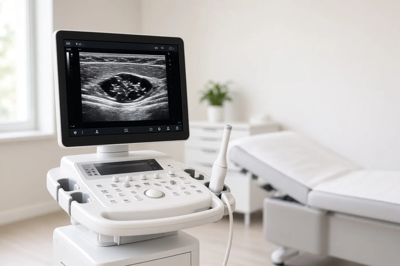
Often, sclerosing adenosis cannot be felt and causes no signs. Sometimes, it forms a small firm lump, which may be tender. Any persistent breast lump or pain should be shown to a doctor.
On the mammogram, it can appear in several ways: a cluster of microcalcifications, that is, very small grains of calcium grouped in the same place; a small mass, with clear or irregular outlines; or an architectural distortion: the usual organisation of the gland looks distorted there.
An ultrasound is often added, and an MRI in some cases. Some of these appearances resemble those of a cancer. The images alone cannot settle the question: the radiologist then most often classifies the image ACR 4 in the BI-RADS system, and offers a biopsy.
Microcalcifications, a mass or a distortion are appearances visible on imaging. Sclerosing adenosis, on the other hand, is a diagnosis made on the tissue, under the microscope. Behind a cluster of microcalcifications, several answers are possible: sclerosing adenosis, another benign lesion, an at-risk lesion or a cancer, for example a ductal carcinoma in situ, which has stayed inside the ducts. Only the microscope can tell the difference.
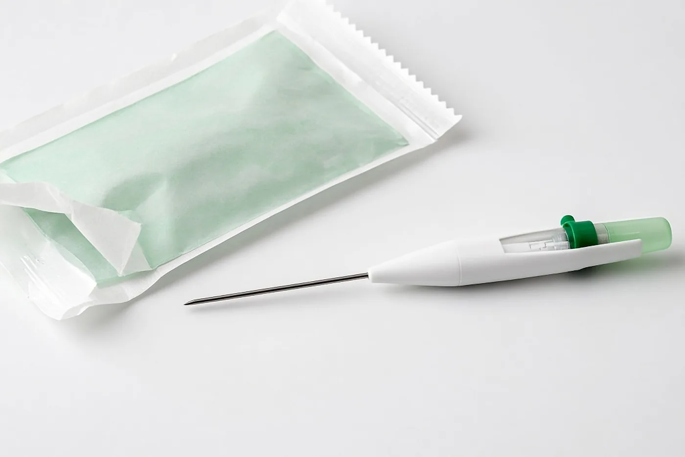
The radiologist takes the sample under local anaesthetic, and you usually go home the same day.
When the image is a cluster of microcalcifications, the recommendations favour a vacuum-assisted biopsy guided by mammogram: a fairly wide needle, connected to suction, collects several fragments of tissue through a single entry point in the skin. For a mass visible on ultrasound, the radiologist most often performs a core biopsy: a finer needle is inserted several times and brings back a small cylinder of tissue at each pass.
A small clip, a metal marker placed where the sample was taken, may be left in. You cannot feel it; it is used to locate the area later, if needed.
Under the microscope too, sclerosing adenosis can look like a cancer. The pathologist then sometimes uses special stains to clear up the doubt, which can slightly lengthen the waiting time for biopsy results.
Once the report has arrived, the radiologist compares the result with what was seen. If the sclerosing adenosis does explain the image, for example microcalcifications found within the area of adenosis on the fragments, this is called concordance. For simple adenosis without atypia, the image can then be reclassified as benign, as ACR 2. If the result does not explain the image, for example a very irregular mass with simple sclerosing adenosis, this is called discordance.
The recommendations provide for these results to be reviewed with the pathologists at a multidisciplinary team meeting (MDT): doctors from several specialties decide together on the course of action. In case of discordance, the adenosis is not enough to explain the image: a new sample is taken, on the decision of the MDT.
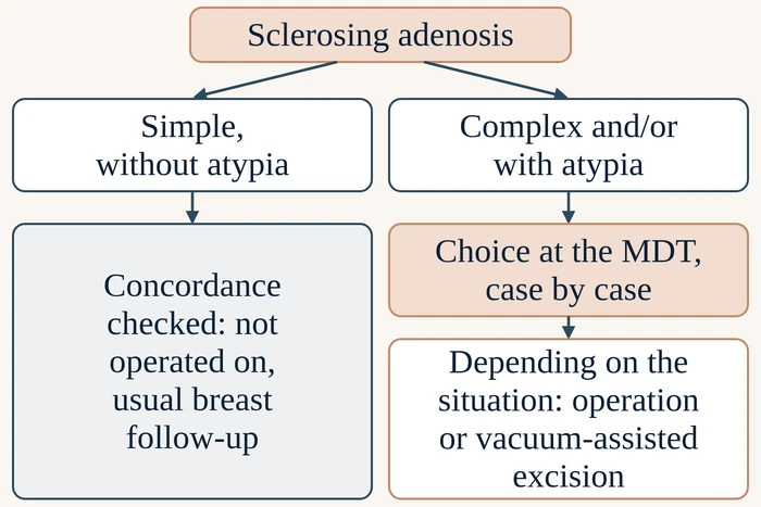
The next step depends on two questions: is the adenosis simple or complex, and did the microscope find atypia? Cases of complex adenosis or adenosis with atypia are examined at the MDT, case by case, to choose between an operation and a vacuum-assisted excision. Radiologists, pathologists, surgeons and oncologists take part: oncologists are the doctors who treat cancer with medicines or radiation.
Once the concordance between the images and the microscope analysis has been checked, simple sclerosing adenosis without atypia is a benign lesion: it is not operated on. Nothing more is required: your usual breast follow-up continues with your gynaecologist or your GP. If a radial scar is also present, its management is explained on its own page.
When the adenosis is complex, or when atypia is present, whether atypical ductal hyperplasia or atypical apocrine adenosis, the area is removed in full, so that it can be examined under the microscope in its entirety. With atypia, the lesion is classified B3 and joins the at-risk lesions. Because the needle takes only a sample of the tissue, a cancer can sometimes lie nearby without having been reached.
Depending on the situation, the removal is done either by an operation, a lumpectomy after preoperative localisation, or by a vacuum-assisted excision, "excision" meaning removal. The choice is made at the MDT, case by case, and Dr Zeitoun discusses it with you.
The operation is performed by Dr Zeitoun. As the area usually cannot be felt by touch, the radiologist places a marker near the clip before the procedure; this step is called preoperative localisation. Dr Zeitoun then removes the tissue around this marker. This is a lumpectomy, that is, the removal of a small part of the gland. The procedure takes place at Clinique Hartmann (Neuilly-sur-Seine).
The vacuum-assisted excision is performed by the radiologist, under local anaesthetic, without going to the operating theatre. The radiologist uses the same needle connected to suction as for the diagnostic sample, but this time removes the whole area visible on imaging.
Complex sclerosing adenosis or sclerosing adenosis with atypia: a consultation with Dr Zeitoun makes it possible to go over your file with all your tests, then present it at the MDT if needed, and prepare the removal of the area together.
REQUEST A CALLBACKBOOK ON DOCTOLIB →Once the area has been removed, by an operation or by a vacuum-assisted excision, all the tissue removed is analysed by the pathologist, and Dr Zeitoun discusses this result with you at a consultation. Most often, only the lesions already known are found. If only benign lesions or atypia are present, a further procedure is most often not needed, even when they reach the edges of the tissue removed. If a cancer is found, your case is presented again at the MDT and the appropriate treatment is explained to you.
For simple adenosis without atypia, once concordance has been checked, your usual breast follow-up continues with your gynaecologist or your GP.
For complex adenosis without atypia, once the area has been removed by an operation or by a vacuum-assisted excision, follow-up takes place with Dr Zeitoun for the first years, then with your gynaecologist or your GP.
With atypia, once the area has been removed, check-ups by mammogram and by ultrasound are spread over ten years: the first years with Dr Zeitoun, then with your gynaecologist or your GP. Atypia makes a future breast cancer more likely, and these check-ups are there to detect it early if it occurs.
Both the operation and the vacuum-assisted excision carry possible complications, for example a haematoma, that is, blood collecting under the skin at the treated site. They are explained to you before the procedure; those of the operation are also listed on the lumpectomy page.

The consultation is for patients whose report mentions complex sclerosing adenosis or sclerosing adenosis with atypia, for whom removal of the area, by an operation or by a vacuum-assisted excision, needs to be organised. It takes place once the biopsy result is available. Dr Zeitoun reviews the images as well as the imaging and pathology reports. He explains the form of sclerosing adenosis found, presents your case at the MDT if needed, then tells you what is being proposed and why.
Bring your images and their disc or access link, the imaging reports, the report on the sample, your tests from previous years and the list of your medicines. Also mention any breast cancers or ovarian cancers in your family.
If a proposal has already been made to you elsewhere, you can ask Dr Zeitoun for a second opinion.
Consultations at the practice, 241 rue du Faubourg Saint-Honoré (Paris 8th), and at Clinique Hartmann (Neuilly-sur-Seine). Surgery takes place at Clinique Hartmann.
Sector 2, not opted into OPTAM, with possible fee supplements. The amount is explained during the consultation and written on a quote before the procedure.
No. It is a benign lesion of the gland, recognised under the microscope. On the images, it can look like a cancer, which is why a biopsy was offered.
Not when it is simple and without atypia: once concordance has been checked, it is not operated on, and your usual breast follow-up continues. When it is complex and/or with atypia, the area is removed, on the decision of the MDT, depending on the situation: either by an operation, a lumpectomy after localisation, or by a vacuum-assisted excision performed with a needle by the radiologist. If a radial scar is also present, its page explains how it is managed.
It moves the result into category B3, that of at-risk lesions, whether it is atypical ductal hyperplasia or atypical apocrine adenosis. The area is then removed, by an operation or by a vacuum-assisted excision, depending on the situation and on the decision of the MDT, to check that no cancer is present next to it. Breast check-ups then continue over ten years: the first years with Dr Zeitoun, then with your gynaecologist or your GP.
No. ACR 4, in the BI-RADS classification, is given by the radiologist looking at the image: it means that the image needs to be sampled. B2 is given by the pathologist examining the tissue: it means that the lesion is benign.
A discordance is examined at the MDT. As the adenosis alone does not explain what the imaging shows, a new sample is taken, on the decision of the MDT.
Not for the simple form: once concordance between the imaging and the microscope has been checked, nothing more is required, and your usual breast follow-up continues with your gynaecologist or your GP. For complex adenosis without atypia, once the area has been removed by an operation or by a vacuum-assisted excision, follow-up takes place with Dr Zeitoun for the first years, then with your gynaecologist or your GP. With atypia, once the area has been removed, check-ups by mammogram and by ultrasound are planned for ten years: the first years with Dr Zeitoun, then with your gynaecologist or your GP.
Complex sclerosing adenosis or sclerosing adenosis with atypia: to review your result and organise the removal of the area, Dr Zeitoun sees you at the Faubourg Saint-Honoré practice or at Clinique Hartmann.
REQUEST A CALLBACKBOOK ON DOCTOLIB →