Home
Breast & breast cancer
Breast cancer Benign breast lesions Prophylactic mastectomy
Breast reconstruction
All techniques Implant reconstruction Latissimus dorsi flap DIEP flap (abdomen) Gracilis flap (thigh) Fat grafting (lipofilling) Flat closure Intimate surgery
Benign gynaecological surgery
Uterus Ovaries and tubes Cervix Vulva and vagina
Gynaecological cancers
Cervical cancer Ovarian cancer Uterine cancer Vulvar cancer Borderline tumours Articles About
Request a callback Book on Doctolib →
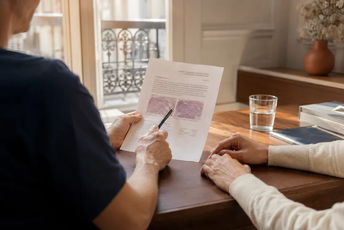
Breast surgeon explaining a breast biopsy report showing an intraductal papilloma to a patient, Paris practice
Benign breast lesions · Decision aid

Breast papilloma: surgery or surveillance?

Your biopsy says "papilloma". It is a benign lesion: it is not a cancer. And yet surgery is often recommended. That is not a contradiction — and the reason deserves to be said plainly: we do not operate on the papilloma, we operate on what may be hiding right beside it. In a small number of cases, an early breast cancer known as DCIS develops in the neighbouring ducts, without the biopsy needle ever having touched it. That is what we are trying not to miss.

📍 Paris 8 · Neuilly Surgery & second opinion 📞 +33 1 58 05 11 24
A biopsy has shown a papilloma?
Review your file and settle the question

Your biopsy report, your imaging, the question of concordance: Dr Zeitoun, breast surgeon, reviews all of it in consultation and tells you what is genuinely indicated — including when the answer is not to operate. Consultations in English, Paris 8 and Clinique Hartmann (Neuilly).

Book on Doctolib Request a callback
THE STARTING POINT

A benign lesion, but a question to settle

The breast contains about fifteen small ducts that carry milk towards the nipple. A papilloma is a small outgrowth that develops inside one of them, arising from its wall.

It is benign. Not a cancer, it invades nothing, it does not spread.

So why talk about surgery? Because of one technical detail of the biopsy. The needle takes two or three small fragments of tissue, about the size of a pencil lead. The area concerned may be one or two centimetres across. So we are looking at a sample, not the whole thing.

And papillomas have one particular feature: right next to them, in the neighbouring ducts, there can be abnormal cells — atypia, and sometimes more. The needle can pass by without seeing them.

What we are really looking for

When excision is recommended, it is not the papilloma that worries us. It is the possibility that a ductal carcinoma in situ — an early breast cancer, also called DCIS — sits in the ducts next door.

DCIS is a cancer that has not yet left the ducts. It cannot be felt, it does not hurt, and on a mammogram — or on an ultrasound — it can look like the papilloma itself. The only way to be certain is to remove the area and examine it in full.

What the guidelines say. The French regional breast reference (SENORIF 2025-2026) reports 10 to 30% of cancers found on the surgical specimen across all B3 lesions. But that figure covers very different situations. For a papilloma without atypia, entirely removed by vacuum-assisted biopsy and fully concordant with the imaging, the residual risk is low — low enough that the guidelines allow radiological follow-up alone in that specific case.

That is the whole logic: excision does not treat a disease, it removes a doubt. And in the great majority of cases, it removes it in the right direction.

That is the whole question. When does this risk justify removing the lesion, and when can we reasonably leave it alone?

UNDERSTANDING YOUR REPORT

That word: "B3"

On your biopsy report there is a letter B followed by a number. It is a code used by every laboratory. It does not say how serious it is: it says what to do next.

GradeWhat the pathologist sawUsual management
B1Normal tissue, or non-representative sampleCheck concordance; often repeat the sampling
B2Clearly benign lesion (fibroadenoma, cyst…)Return to routine surveillance
B3Lesion of uncertain malignant potential: papilloma, atypical ductal hyperplasia, radial scar, phyllodes tumourMultidisciplinary discussion: further excision or surveillance
B4Suspicious, without certaintyFurther sampling or excision
B5Malignant (B5a in situ, B5b invasive)Oncological management

The mistake almost every patient makes: reading "B3" as "slightly cancerous". It is nothing of the sort. B3 means: what we sampled is benign, but that sample is not enough to answer for the whole area. It is about certainty, not severity.

Breast biopsy report marked B3, a lesion of uncertain malignant potential, on the breast surgeon's desk
On your report, “B3” does not mean “slightly cancerous”: it means the biopsy alone is not enough to reach a conclusion.

Why the papilloma belongs to this group

Two reasons, one anatomical, one statistical.

Anatomical. A papilloma is both bulky and heterogeneous. It fills the lumen of a duct, sometimes extending several centimetres along it, and its immediate surroundings may harbour foci of atypia. Sampling a few fragments of such a lesion means probing the ground, not mapping it.

Statistical. When papillomas diagnosed on biopsy were later removed surgically, a DCIS — or more rarely an already invasive cancer — was sometimes found alongside. Not often. But often enough that no serious team ignores the question.

That figure is what drives the whole decision, and it varies considerably between situations — which is the subject of the next section.

Does your report say "B3"?

That is the moment to see a breast surgeon. Dr Zeitoun reviews the report and your imaging, and tells you whether surgery is needed.

THE WORD THAT CHANGES EVERYTHING

Look for the word "atypia"

If you take only one thing from your report, take this. Look for the word "atypia" or "atypical". Whether it is there or not separates two situations that carry neither the same risk nor the same management.

01

Papilloma without atypia

The cells are regular and normally organised. The risk of finding a DCIS is then low. This is the only situation where radiological follow-up alone is endorsed by the guidelines — provided the vacuum-assisted biopsy removed everything and the imaging concords.

02

Papilloma with atypia

Some cells have lost their regularity. The risk of finding a DCIS or a cancer alongside rises sharply. The guidelines are explicit: for a papilloma with atypia, surgical excision is the standard treatment, whatever the sampling method.

03

Central or multiple

Whether it is central (behind the nipple) or peripheral changes the approach: a central papilloma, too close to the nipple-areola complex, does not lend itself to vacuum-assisted biopsy and calls for surgery. Multiple papillomas also carry a higher risk of associated atypia.

Patient reading an information leaflet in the waiting room of a breast surgery practice in Paris
Between the biopsy and the decision, the waiting is often the hardest part. It shortens as soon as a surgeon goes through the file with you.

A useful word about the figures. These percentages vary from one publication to another, sometimes twofold. That is not a flaw in the literature: it reflects different populations, different biopsy techniques, and above all different selection criteria. An isolated figure, taken out of context, tells you nothing about your own case. What matters is the direction it points in — and that is consistent across studies: atypia raises the risk, complete excision lowers it.

THE DECISION

What tips the balance

The decision is never taken on a single item. It comes from a body of arguments, discussed at a multidisciplinary team meeting bringing together surgeon, radiologist and pathologist. Here are the criteria actually used.

CriterionPoints towards excisionAllows surveillance to be discussed
AtypiaPresentAbsent
SymptomsBloodstained discharge, palpable lumpNo symptoms, incidental finding
Lesion size (15 mm threshold)15 mm or more — beyond that, vacuum-assisted biopsy no longer removes it allUnder 15 mm, the threshold used in the guidelines
Sampling methodCore biopsy alone (limited material)Vacuum-assisted biopsy that removed the whole target
Radiology–pathology concordanceDiscordance between image and resultFull concordance
Number of lesionsMultiple, peripheral papillomasSingle, behind the nipple
Personal contextBreast cancer already treated, family historyNo particular risk factor
Your ageA younger woman — many years of surveillance still aheadAfter the menopause, with no risk factor
How you feel about itThe uncertainty weighs on you, you want it settledYou are comfortable with regular follow-up

Concordance between image and result

It is the most technical of the seven, and the most decisive. Radiology–pathology concordance means bringing two viewpoints together: what the radiologist saw on the image, and what the pathologist found in the sample. If the radiologist described a one-centimetre mass and the laboratory describes a one-centimetre papilloma, the story is coherent — that is concordance.

But if the radiologist described an image graded BI-RADS 4 or 5 and the sample yields only a small ordinary papilloma, there is discordance. What worried the radiologist was probably not what was sampled. In that case the benign result is not reassuring: further work is needed. This check is done systematically, and it is the single best protection against error.

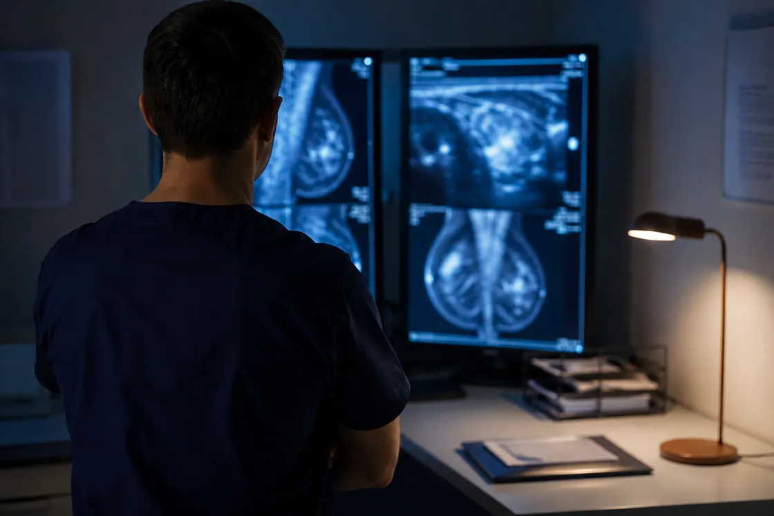
Breast radiologist reviewing a mammogram and a breast ultrasound on reporting monitors
Concordance: does what the radiologist saw on the image match what the laboratory found? It is the single most decisive criterion.

The three real options

Vacuum-assisted excision. Performed by the radiologist, under local anaesthetic, with a large-bore vacuum-assisted needle that removes the lesion in successive fragments. No theatre, no scar, no general anaesthetic. Suited to small lesions without atypia. Its limit: removal is not always complete, and the tissue is fragmented, which makes the analysis slightly less precise.
Microdochectomy. Surgical excision of the affected duct, in one piece, in theatre as a day case. This is the reference option when there is atypia, a discharge, a bulky lesion or a discordance. It gives the most reliable analysis, at the cost of a scar.
Radiological follow-up. The guidelines allow it in one precise situation: a papilloma without atypia, removed in full by vacuum-assisted biopsy, with concordant imaging. Those conditions must be met together. And it is not "doing nothing": it is a programme of appointments and imaging at fixed intervals over several years, which you must be prepared to follow.
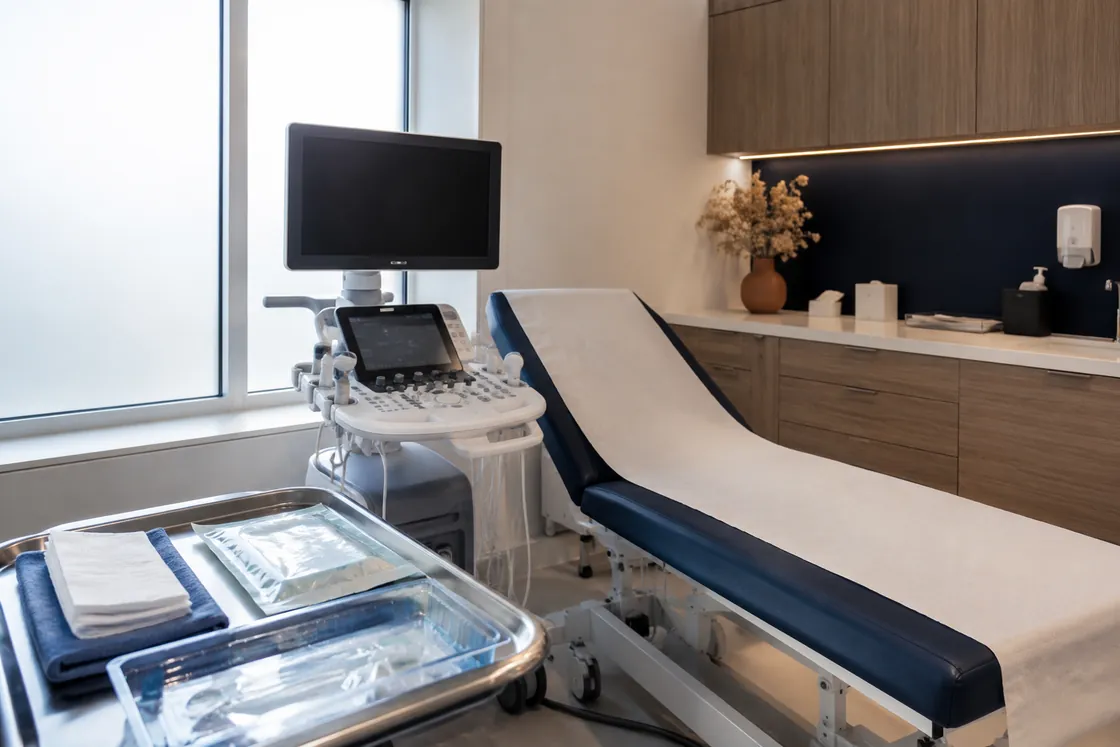
Interventional ultrasound room equipped for a vacuum-assisted breast biopsy
Vacuum-assisted excision is carried out in the ultrasound room, under local anaesthetic, with no theatre and no scar.

When we remove it

Beyond the table, four situations tip the balance towards excision — and they are the ones seen most often in clinic. None of them decides on its own: it is for the surgeon to weigh the advantages and drawbacks of each option, with you, after discussion.

You are young. The longer surveillance has to run, the more checks accumulate — and each one is another occasion for doubt. Removing the lesion once settles the question for good. The surgeon weighs that against the drawbacks of the procedure.
You have a family history. A mother, a sister or an aunt affected by breast cancer changes the equation. The background is no longer neutral, and tolerance for doubt has to be lower. Where the family history is strong, a genetics referral is discussed alongside — and where a mutation is confirmed, risk-reducing mastectomy becomes a separate discussion in its own right.
You have symptoms. Bloodstained discharge from a single pore, a lump you can feel: these are not incidental findings. The lesion is making itself known — and such signs will not go away under surveillance.
The uncertainty weighs on you. This is a criterion in its own right, and there is nothing illegitimate about it. Living for years with a doubt about a possible early cancer is not neutral: sleep, anxiety before every check, difficulty planning ahead. If you know you will not cope well with it, say so. It counts as much as the other criteria, and it is often what settles the question.

Why a surgical opinion changes the decision

The seven criteria are not read in isolation, they are weighed. A 9 mm papilloma without atypia but with bloodstained discharge is not managed like a 9 mm papilloma found by chance — yet both occupy the same row of the table.

The guidelines also note that agreement between pathologists is lower for B3 lesions than for cancers, and recommend having the slides reviewed by an expert where there is doubt. That is exactly what a second opinion allows you to trigger.

What a surgical consultation actually adds: going through the report with you, comparing it against your imaging, checking that what worried the radiologist was in fact sampled, and giving a clear answer — operate, monitor, or sample again. Including when the answer is to do nothing.

Multidisciplinary breast team meeting reviewing a B3 breast lesion case
The decision is never taken alone: surgeon, radiologist and pathologist review the case together.

Dr Zeitoun operates on these lesions every week at Clinique Hartmann in Neuilly-sur-Seine, as day surgery, and consults in English. He also sees patients for a second opinion — and how to choose a breast surgeon sets out the criteria that matter when excision has already been proposed elsewhere and you would like to discuss it before deciding.

THE PROCEDURE

How the operation goes

In French the operation is called a pyramidectomie, after the shape of the tissue removed: a pyramid whose tip reaches the nipple and whose base extends into the breast, following the course of the affected duct.

30–45minutesUsual duration of the procedure itself.
D0Home the same dayDay surgery: admission in the morning, home by the end of the day.
3–4cm scarIncision placed at the edge of the areola, in the natural skin transition.
3–7daysReturn to everyday activities; sport waits two to three weeks.
10–15daysTime to the final histology result, given in consultation.

What we remove, and why

The aim is not to remove "the papilloma" but the duct that contains it, along its useful length. Removing the lesion alone would leave the surrounding tissue in place — precisely where the atypia we are trying to rule out may sit.

If the lesion is not palpable, pre-operative localisation is carried out the day before or the same morning by the radiologist: a wire, a magnetic seed or ultrasound guidance depending on the case. When a discharge is present, the responsible duct can be identified directly during the operation. The incision follows the edge of the areola, the dissection runs along the duct from the nipple, and the specimen is removed en bloc — it is this single-piece removal that allows the pathologist to assess the margins and the periphery properly.

General anaesthetic, with a mandatory anaesthetic consultation at least 48 hours beforehand. Closure with absorbable sutures, simple dressing. The detail of recovery — pain, scar, return to activity — is set out on a dedicated page.

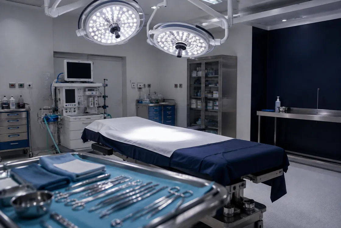
Day-surgery breast operating theatre prepared at Clinique Hartmann, Neuilly-sur-Seine
Removing a papilloma is a short procedure, carried out as day surgery at Clinique Hartmann (Neuilly-sur-Seine).

What can happen

They are uncommon, but they exist and should be stated beforehand, not discovered afterwards.

ComplicationFrequencyManagement
Haematoma in the surgical bedAbout 2%Usually simple monitoring; return to theatre is exceptional
Wound infection1 to 2%Local care, antibiotics if needed
Altered nipple sensationCommon, transientUsually settles within a few months; may partly persist
Nipple retraction or dimplingRareDepends on the extent of excision; correction possible later
Difficulty breastfeeding on the operated sideRare if one duct removedTo be anticipated in consultation if pregnancy is planned

Take stock before deciding

A younger woman, a family history, bloodstained discharge, or simply wanting to be done with the doubt: in most cases excision is the right answer. A consultation confirms it and allows surgery to be arranged quickly.

AFTERWARDS

The result, and what comes next

The specimen goes to the histopathology laboratory, where it is examined in full. The final result comes back in ten to fifteen days and is given to you in consultation, never by letter alone. Three possibilities:

Benign papilloma confirmed, no atypia. The most frequent outcome. The question is closed. You return to normal surveillance, adapted to your age and history.
Atypia found on the specimen. The lesion remains benign, but it places you in the group of at-risk lesions. It changes nothing about what has been done, but it does change follow-up: closer imaging, and sometimes a genetics referral depending on family history.
Carcinoma in situ or invasive cancer found. This is the minority situation the whole approach was designed not to miss. The case then goes to the multidisciplinary meeting and follows the breast cancer pathway. Found at this point, it is found very early: treatment then usually rests on a lumpectomy, sometimes combined with an oncoplastic technique to preserve the shape of the breast; mastectomy is reserved for extensive lesions.

A reassuring point that often goes unsaid. If the analysis shows the lesion was not removed with entirely clear margins, there is no re-excision to plan. The guidelines state it plainly: for a B3 lesion, incomplete excision does not warrant further surgery. Only finding an associated cancer changes the approach.

And in the long run?

A single papilloma without atypia raises the later risk of breast cancer little if at all. You return to the surveillance rhythm appropriate for your age — screening mammography where a programme applies, or a personalised schedule if you have a relevant history.

A papilloma with atypia, or multiple papillomas, do place you in a somewhat higher-risk group. Follow-up then becomes more structured, often annual, and, depending on your breast density, sometimes with a breast MRI depending on context — the breast MRI page explains when it is indicated. This increase in risk applies to both breasts, not only the operated one: it is an important point, and it is often poorly conveyed.

Finally, recurrence at the same site is possible but uncommon after complete excision. A new discharge, a new lump or a new imaging finding should be reported without waiting for the next appointment.

Breast surgeon giving the final histopathology result in consultation after removal of a papilloma
The final result is always given in consultation, never by letter alone.

Your questions

The questions that come up most often after a biopsy showing a papilloma. If yours is not here, ask it at your appointment — or ask Sophie, the site assistant, bottom right.

My papilloma is benign — why am I still being offered surgery?

Because we do not operate on the papilloma: we operate on what may be beside it. A biopsy takes only two or three fragments of a much larger area. In the neighbouring ducts there may be a DCIS — an early breast cancer — that the needle never touched. It cannot be felt and does not hurt. Removing the area allows it to be examined in full and the doubt to be settled for good. In the great majority of cases there is nothing.

What is a "B3" lesion?

Breast biopsy results are graded B1 to B5. B3 covers lesions of uncertain malignant potential: the papilloma is a leading example, along with atypical ductal hyperplasia, radial scar and phyllodes tumour. B3 does not mean "slightly cancerous": it means the biopsy result alone is not enough to close the question.

Can we simply keep an eye on it?

It is uncommon. Every condition must be met at once: no atypia — check the wording on your pathology report — a small lesion, no symptoms, a vacuum-assisted biopsy that removed the whole target, imaging in complete agreement with the pathology, no family history — and an age that lends itself to it. In practice most papillomas are removed, and rightly so. A younger woman, a patient with a family history, or one with bloodstained nipple discharge, all warrant excision. And if the uncertainty weighs on you, that alone is reason enough.

What difference does atypia make?

It changes the management. Without atypia, the risk of finding a cancer on the excision specimen is low, of the order of a few per cent. With atypia it is markedly higher — studies most often place it between one case in five and one in three. In that case excision is recommended, and surveillance alone is no longer reasonable.

Vacuum-assisted excision or surgery: what is the difference?

Vacuum-assisted excision is done by the radiologist, under local anaesthetic, with a large-bore needle that removes the lesion in fragments: no theatre, no scar, but it suits small lesions and does not always guarantee complete removal. Microdochectomy is surgery in theatre under general anaesthetic, as a day case: it removes the affected duct in one piece, allowing a more reliable analysis, at the cost of a scar at the edge of the areola.

Will the operation leave a visible scar?

The incision is placed at the edge of the areola, on the boundary between coloured and pale skin, where it blends into the natural transition. It usually measures 3 to 4 centimetres. Red for a few weeks, it fades gradually over twelve to eighteen months. Protecting it from the sun during the first year noticeably improves the result.

Will I lose sensation in the nipple?

Altered sensation in the nipple and areola is common in the first weeks: the dissection passes close to the small nerves supplying them. It usually settles within a few months. An area of reduced sensation may persist, especially if the excision had to be extensive. This is worth discussing before the operation.

Will I be able to breastfeed afterwards?

Most often yes. Microdochectomy removes only one duct or a small group of ducts, out of the fifteen or so in a breast, and the other breast is unaffected. Milk supply on the operated side may be reduced. If several ducts must be removed, feeding from that side may become difficult: if you are planning a pregnancy, say so beforehand — it forms part of the decision.

Does a papilloma raise my risk of breast cancer later?

A single papilloma without atypia raises later risk little if at all. A papilloma with atypia, or multiple peripheral papillomas, are however regarded as at-risk lesions: they warrant structured follow-up with regular imaging, and sometimes a genetics referral if the family history suggests it.

Should I ask for a second opinion?

It is entirely reasonable, and for a B3 lesion it is often useful: the decision rests on a body of arguments that different teams may weigh differently. A second surgical opinion allows the report, the imaging and the question of concordance to be reviewed. It does not delay treatment — there is no urgency in operating on a papilloma.

Read also

To take your understanding of the diagnosis and its management further.

SOURCES & REFERENCES

Sources

This article draws on French and European guidance on B3 breast lesions.

SFSPM / SENORIF — 2025-2026 recommendations on the management of B3 breast lesions (sfspm.fr).
Rageth CJ et al.Second and Third International Consensus Conference on lesions of uncertain malignant potential in the breast (B3 lesions), Breast Cancer Research and Treatment.
Rubio IT et al. — European guidelines on the management of B3 breast lesions, European Journal of Surgical Oncology, 2024.
Institut National du Cancer (INCa) — Benign and at-risk breast lesions (e-cancer.fr).
ACR BI-RADS Atlas, 5th edition — classification of breast imaging findings and the principle of radiology–pathology concordance.

Written and medically reviewed by Dr Jérémie Zeitoun, breast surgeon. Last reviewed: 1 August 2026. This content is informational and does not replace a medical consultation.

A papilloma diagnosed: operate, or monitor?

Has a biopsy shown a papilloma? Has excision been proposed and you would like a second opinion before deciding? Bring your report and your imaging — see how to prepare for your first consultation. Dr Jérémie Zeitoun, breast surgeon trained at Gustave Roussy and Institut Curie, consults in English at his practice in the 8th arrondissement of Paris and operates as day surgery at Clinique Hartmann in Neuilly-sur-Seine. See also choosing a breast cancer surgeon in Paris.

FR EN