A redness, a scab or scaling of the nipple that looks like eczema but will not heal: this is how Paget's disease of the nipple can present — a rare form of breast cancer. Its hallmark: this visible skin change is most often the sign of a cancer sitting beneath the nipple. The good news is that it is diagnosed with a simple procedure and treated well when caught in time. Here, from the surgeon's side, is what you need to know.
You have a biopsy result and an imaging work-up: Dr Zeitoun reviews them, sets the surgical strategy — conservation or mastectomy — and operates at the Clinique Hartmann in Neuilly. Also for a second opinion. No biopsy yet? here is what to do.
Most nipple changes are benign; the aim of a consultation is simply not to miss a diagnosis that is treated well when made early.
It looks like nipple eczema: a redness, some scaling, small crusts, sometimes itching. You apply a cream, it improves a little, then it comes back. What sets Paget's disease of the nipple apart from simple irritation is precisely this persistence: a change that will not heal as it should.
This condition is a rare form of breast cancer (about 1 to 4% of cases). Its hallmark comes down to one image: it is usually the “visible part” of a cancer sitting deeper in the breast — most often a ductal carcinoma in situ, sometimes an invasive cancer. The skin lesion is the signal; it is what hides beneath that guides treatment.
This article explains, from the surgeon's point of view: which signs should alert you, how the diagnosis is made, what is looked for beneath the nipple, and which surgery to offer — conserving or mastectomy. It draws on the SÉNORIF 2025-2026 guideline, the INCa and ESMO, and complements the breast cancer pillar page.

Paget's disease of the nipple is a rare form of breast cancer (1 to 4% of cases) in which cancer cells invade the skin of the nipple and areola. It looks like eczema that will not heal, on one side only, despite creams. In close to 9 out of 10 cases a breast cancer is present underneath. The diagnosis rests on a biopsy of the nipple skin.
No single feature settles it. It is the combination — and above all persistence on one side — that makes a biopsy necessary. See also: breast cancer, breast lump, nipple discharge.
The name comes from the English surgeon Sir James Paget, who described it in the 19th century. Behind a change in the skin of the nipple lies, almost always, a disease of the breast.
In Paget's disease, distinctive cancer cells — Paget cells — migrate into the skin of the nipple and areola (the nipple-areola complex). This is what gives the “eczema” appearance.
But these cells almost always come from a cancer sitting inside the breast, beneath the nipple. Understanding Paget's disease therefore means looking at two levels: the skin you can see, and the lesion looked for beneath it.
It accounts for about 1 to 4% of breast cancers. It mainly affects women after 50, but can, more rarely, occur in men.
In around 9 out of 10 cases, an underlying cancer is found: most often a ductal carcinoma in situ, sometimes an invasive cancer.
It affects a single breast and starts at the nipple before spreading to the areola — unlike an ordinary eczema, often on both sides and fluctuating.
The key point: Paget's disease of the nipple (the breast) must not be confused with extramammary Paget disease (vulva, groin, armpit) or Paget's disease of bone, which are entirely different conditions sharing the same name. This article concerns the breast only.
The manifestations resemble those of an ordinary skin condition. What matters is not any single sign, but their persistence on one side despite topical treatment.
The nipple becomes red, scaly or crusty, the skin peels or thickens. It may look weepy. These signs start at the nipple and then spread to the areola — never the other way round.
Itching, a burning or tingling sensation are common. They may precede the visible skin changes and suggest, wrongly, a simple irritation.
A serous discharge or slight bleeding from the nipple may occur. A nipple discharge, especially one-sided and spontaneous, always deserves an opinion.
The nipple may flatten or retract. Sometimes a lump is felt near the nipple — a sign pointing to an underlying lesion to look for.
The simple rule to remember: an ordinary nipple eczema clears with creams within two to three weeks. A nipple change that persists, recurs or worsens despite proper topical treatment should be checked — without panic, but without letting it drift. An examination and, if needed, a biopsy settle the question quickly.
Faced with a nipple lesion that persists, one simple procedure gives certainty: take a small fragment of the nipple skin and analyse it under the microscope.
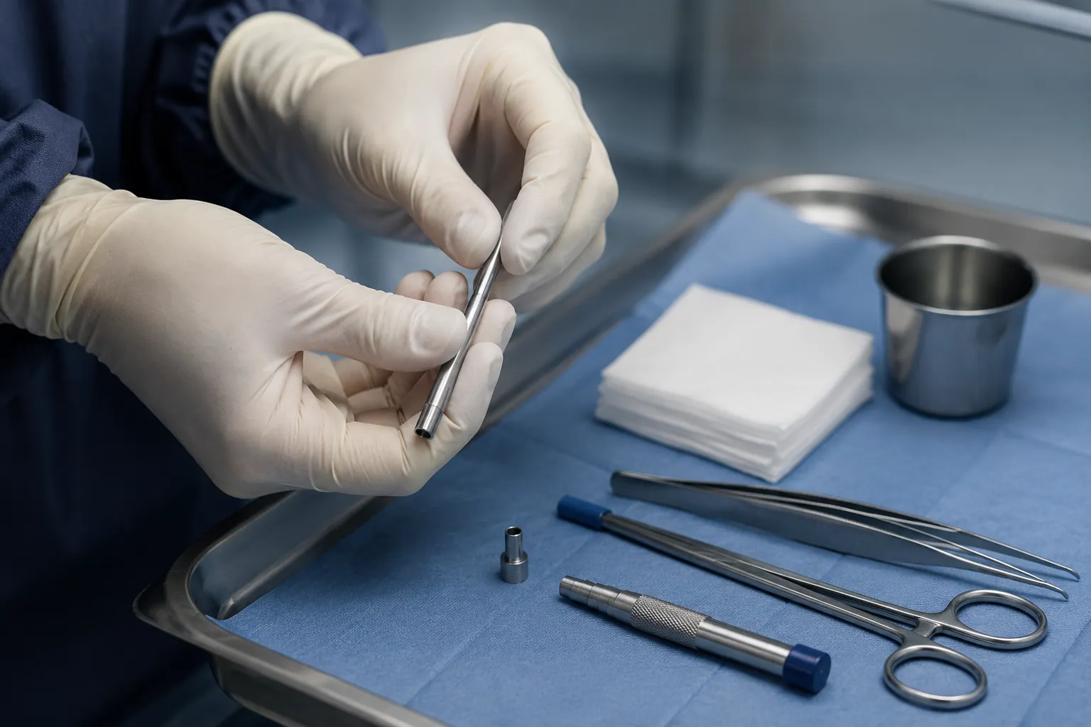
The diagnosis rests on a biopsy of the nipple skin (a skin biopsy, often taken with a “punch”, a small circular tool), under local anaesthetic. Microscopic analysis (pathology) shows the Paget cells and confirms the diagnosis. It is a quick, well-tolerated procedure, which may be completed by a breast biopsy if imaging shows a deeper abnormality.
A mammogram looks for microcalcifications or a mass. It is essential, but can be normal: a normal mammogram does not rule out the disease. This is especially true with dense breasts, where the images are harder to read. If a finding is classified BI-RADS 4 or 5, what happens next is set out in our article on BI-RADS 4 and 5 results.
The breast ultrasound complements the mammogram, particularly to explore the area behind the nipple and the armpit, and to guide a biopsy.
The breast MRI is very useful when the mammogram is normal: it detects and measures a lesion not otherwise visible, and defines its extent before surgery.
Good to know: the diagnosis of Paget's disease is sometimes made late, because the lesion is taken for eczema for months. That is why the message is simple: a nipple lesion that will not heal should be biopsied. The result immediately guides what comes next — usually within a few days to two weeks, as explained in our article on how long breast biopsy results take.
This is the most common situation, and the order of steps matters. The nipple biopsy comes before the surgical consultation: it is what tells you whether this is Paget's disease or an ordinary skin condition, and therefore whether surgery is called for at all.
It is with that result in hand that the surgical consultation becomes truly useful. Dr Zeitoun reviews the pathology report and the imaging, presents the file at the MDT, and sets the strategy: breast-conserving surgery or mastectomy, with or without a sentinel node biopsy, and the reconstruction options.
If the biopsy could not be arranged, or if doubt persists despite a first sample, Dr Zeitoun can perform it himself in clinic. And if a diagnosis has already been made elsewhere, a second opinion before surgery is entirely legitimate.
Dr Zeitoun reviews your biopsy report and imaging, sets the surgical strategy and operates — at the Clinique Hartmann in Neuilly.
The skin lesion signals the disease; it is the deeper lesion that determines its seriousness and treatment. The whole work-up aims to find and characterise it.
Most often, a ductal carcinoma in situ (DCIS) is found: a “contained” cancer, still inside the ducts, non-invasive. It is the earliest form, with a good outlook when treated. It is the same DCIS that we set out to rule out when a biopsy shows an intraductal papilloma.
In a proportion of cases, the underlying lesion is an invasive cancer. The risk is higher when a lump is palpable: one more reason to consult early, before a lump appears.
The work-up establishes whether the disease is localised (a single area, near the nipple) or multifocal/extensive. This distinction is decisive: it points towards breast-conserving surgery or a mastectomy.
On the samples, the grade of the lesion is specified. Hormone receptors and HER2 are tested only if a cancer is actually found beneath the nipple — and it is then the underlying cancer, not Paget's disease itself, that is characterised. This profile therefore does not apply to every patient: it serves to decide any additional treatment after surgery, at the MDT.
Bear in mind: when there is no palpable lump, the underlying lesion is most often an in-situ cancer, with a very good outlook. The presence of a lump raises more concern about an invasive component — hence the importance of imaging and biopsy to map everything before operating.
Surgery is the main treatment. One point is common to every option: the nipple and areola (where the Paget cells sit) must always be removed. This choice between conserving and removing follows the same rules as for other breast cancers — set out in lumpectomy or mastectomy: how the choice is made. The rest depends on what has been found beneath.
For localised disease with achievable clear margins, the nipple-areola complex and the affected area are removed (breast-conserving surgery), followed by radiotherapy to the breast. A validated alternative to mastectomy in selected cases.
If the disease is extensive, multifocal or if clear margins cannot be achieved, mastectomy remains the safest solution. A reconstruction — immediate or delayed — is possible within the same pathway.
For an invasive cancer, or when a mastectomy is performed, the armpit is assessed with the sentinel node — a targeted procedure that often avoids full clearance and its risk of lymphoedema. An isolated in-situ cancer does not always require this step.
The removed nipple and areola can be reconstructed at a second stage (a small plasty, reconstruction, areola tattoo). Oncoplastic surgery helps preserve a fine breast shape after conservation.
Always a shared decision: the choice between conservation and mastectomy is made at a multidisciplinary team meeting (MDT), according to the extent of the disease, the expected quality of the margins and your wishes. The recovery is generally straightforward.
It is the full analysis of the operative specimen that dictates what follows. The additional treatment is not aimed at the “skin” of Paget, but at the cancer found beneath.
After breast-conserving surgery, radiotherapy to the breast is usual: it completes the surgery and reduces the risk of local recurrence.
Depending on the underlying cancer: hormone therapy if the tumour is hormone-sensitive, targeted therapies or chemotherapy for an invasive cancer.
Regular follow-up (clinical and imaging) is set up. Testing for a genetic predisposition (hereditary breast-ovarian risk) is offered according to family history.
On prognosis: it depends above all on the lesion found beneath the nipple — an in-situ cancer, with no palpable lump, has a very good outlook; an invasive component, or node involvement, is managed differently. The outlook is therefore individual: it is discussed with your team, and no figure can be given from reading an article alone.
From the first examination of the nipple to surgery and follow-up, Dr Zeitoun coordinates each step with the multidisciplinary team.
Most often you arrive with a biopsy result already available. Dr Zeitoun reviews the pathology report and the images, examines the nipple, and completes the work-up if needed. If the biopsy has not yet been done, he can perform it. Prepare your visit with the consultation checklist, and see how to choose a breast cancer surgeon in Paris.
He establishes what lies beneath the nipple, presents your file at the MDT, and offers you the most suitable strategy: conservation where possible, mastectomy where preferable.
He performs the excision of the nipple-areola complex and the necessary node procedure, thinking from the outset about the aesthetic result and the options for reconstruction.
Consultations at the practice in the 8th arrondissement of Paris (241 rue du Faubourg Saint-Honoré), operations at the Clinique Hartmann in Neuilly-sur-Seine. Sector 2 (non-OPTAM): a quote is provided before any procedure.
It is a rare form of breast cancer (about 1 to 4% of cases) that shows up as a change in the skin of the nipple and areola. Distinctive cancer cells — Paget cells — are present in the skin of the nipple. In the vast majority of cases, this skin change is the visible sign of a cancer sitting deeper in the breast. It should not be mistaken for a simple eczema. Diagnosis and management are decided at a multidisciplinary team meeting (MDT).
No — most nipple changes are benign (eczema, irritation, thrush). But there is a simple rule of thumb: an ordinary eczema clears with creams within two to three weeks. A nipple change that persists, comes back or worsens despite proper topical treatment should be checked, because it may reveal Paget's disease. The point is not to panic, but not to let it drift: a lesion that will not heal deserves an opinion.
Almost always. In around 9 out of 10 cases, an underlying cancer is found in the breast: most often a ductal carcinoma in situ (a “contained”, non-invasive cancer), sometimes an invasive cancer. This is exactly why the work-up systematically looks for what lies beneath the nipple: it is that deeper lesion that determines treatment.
The key test is a biopsy of the nipple skin (a skin biopsy, often a “punch” biopsy), done under local anaesthetic: it shows the Paget cells. It is completed by imaging — mammogram, ultrasound and often breast MRI — to look for and locate any underlying cancer. Microscopic analysis (pathology) confirms the diagnosis.
Yes. In some patients the mammogram shows no abnormality and there is no palpable lump. A normal mammogram therefore does not rule out the disease: it is the nipple biopsy that makes the diagnosis. Breast MRI is particularly useful in these situations, to detect and measure a lesion not visible on the other tests.
No, not always. Historically, mastectomy was the rule. Today, for localised disease and when clear margins can be achieved, breast-conserving surgery is possible: the nipple-areola complex and the affected area are removed, followed by radiotherapy to the breast. The choice depends on the extent of the underlying disease and is decided with you, at the MDT.
In limited forms, yes — provided the nipple and areola (where the Paget cells sit) are removed completely with clear margins, followed by radiotherapy. Breast-conserving surgery can be combined with oncoplastic techniques, and the nipple can be reconstructed later. If the disease is extensive or multifocal, mastectomy remains preferable.
It depends on what is found beneath the nipple. If there is only a carcinoma in situ, node assessment is not always needed. In the case of an invasive cancer, or when a mastectomy is performed, the armpit is assessed using the sentinel node technique — a targeted procedure that often avoids full clearance and its risk of lymphoedema.
They depend entirely on the lesion found beneath the nipple, specified by the analysis of the operative specimen. After breast-conserving surgery, radiotherapy is usual. Depending on the underlying cancer, hormone therapy (if the tumour is sensitive to it), targeted therapies or chemotherapy may be offered. All these decisions are made at the MDT.
It mainly affects women after 50, but it can, more rarely, occur in men, like male breast cancer. In men as in women, a nipple lesion that persists or bleeds should never be ignored: it warrants a consultation and, if needed, a biopsy.
With the diagnosis made, the next step is a breast surgery consultation with your results. Bring the pathology report and all of your imaging (mammogram, ultrasound, MRI if one was performed). The surgeon reviews the file, completes the work-up if needed, presents your case at the multidisciplinary team meeting (MDT), and then proposes a strategy: breast-conserving surgery with excision of the nipple-areola complex, or a mastectomy, with or without a sentinel node biopsy. Reconstruction options are discussed from that first consultation.
Yes, and it is common. A second opinion is your right, it offends no one, and it does not cost you time: Paget's disease progresses slowly, and a few days of reflection do not change the outlook. It is particularly useful when a mastectomy is proposed while you are wondering about keeping the breast, or when you want to discuss reconstruction options. Bring the biopsy report, the images on CD or via a download link, and the MDT record if one already exists.
To go further into the diagnosis, surgery and treatments of breast cancer.
The reference page: diagnosis, surgery and treatments of breast cancer, step by step.
UnderstandThe “contained” cancer most often found beneath Paget's disease: what it changes.
A symptomWhen a discharge should be assessed, and what is looked for.
The diagnosisHow a biopsy is done, and what it contributes to the diagnosis.
SurgeryWhen it is needed, how it is performed and the reconstruction options.
SurgeryChecking the armpit without needless clearance, for an invasive cancer.
Your nipple biopsy has shown Paget's disease, or you would like a second opinion before surgery: Dr Jérémie Zeitoun reviews your file, presents your case at the MDT and agrees the surgical strategy with you. Practice in the 8th arrondissement of Paris, operations at the Clinique Hartmann in Neuilly-sur-Seine. Bring your imaging and, where relevant, biopsy reports.
This article is for information and does not replace a medical consultation; the diagnosis and treatment strategy are established with the multidisciplinary team.