


This article is based in particular on the SENORIF 2025-2026 guidelines.
You have just learned that a local recurrence is possible, or already confirmed?
Bring the biopsy report, the images, the report of the first operation and those of the treatments already received. Dr Jérémie Zeitoun, surgical oncologist and breast surgeon, sees patients at the practice at 241 rue du Faubourg Saint-Honoré (Paris 8th) and at Clinique Hartmann (Neuilly-sur-Seine). Surgery takes place at Clinique Hartmann.
A teleconsultation is possible to review the file before an in-person consultation. You can book an appointment on Doctolib or request a callback from the secretariat.
Local recurrence, regional recurrence or new cancer
These three situations are not treated in the same way. The work-up serves first of all to tell them apart.
Local recurrence
A local recurrence is the reappearance of cancer cells in the breast that has already been treated, most often near the scar or in the bed of the first tumour. After a mastectomy, it is located on the chest, in the skin or the scar.
It can be invasive, meaning that the cells have crossed the basement membrane of the ducts or lobules, or it can be a ductal carcinoma in situ, meaning that the cells remain inside the ducts or lobules. This distinction, made on the biopsy and then on the surgical specimen, changes the work-up and the treatment.
A local recurrence is not a metastasis. A metastasis means that cells have settled in another organ, for example a bone, the liver or the lung. An isolated local recurrence remains a disease of the breast or the chest, most often treated with the aim of cure.
Regional recurrence
A regional recurrence is often in the axillary lymph nodes, that is, located in the lymph nodes of the armpit. When the involvement is supraclavicular or infraclavicular, above or below the collarbone, or internal mammary, behind the breastbone, it is rather called a locoregional recurrence. A lump under the armpit after breast cancer is not necessarily a recurrence: a lymph node can react to an infection or a vaccine.
Only the examination, the ultrasound and, if needed, a biopsy can tell. When lymph nodes are involved, the treatment generally combines a surgical procedure, often radiotherapy of the area, and in some cases chemotherapy, sometimes combined with immunotherapy or targeted therapy.
A new cancer

A new cancer, distinct from the first, can occur in the other breast or in the same breast. It then differs in its location and/or its characteristics: the type of cells seen under the microscope, known as the histological type, and the receptors (for oestrogen, for progesterone, or the HER2 protein). It is distinguished from a true recurrence.
A new cancer in the other breast is treated like a first cancer, with its own work-up. The distinction relies on the site, the type of cells, the markers and the time since the first treatment. In case of doubt, the guidelines reason as for a recurrence.
The signs that should prompt a consultation
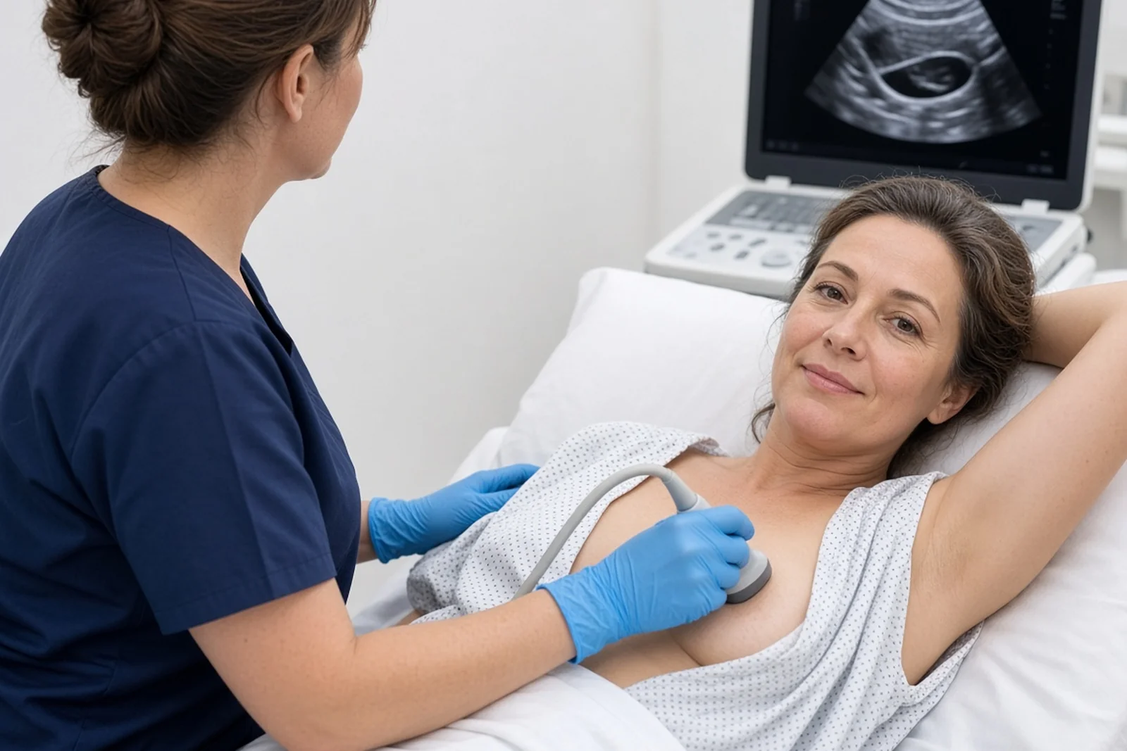
Many local recurrences are found on the yearly mammogram and ultrasound, sometimes in the form of a cluster of microcalcifications, a distortion or a nodule near the scar. Others are revealed by a sign that you notice yourself.
See a doctor without waiting for the next scheduled visit in case of a new lump in the treated breast, near the scar or in the other breast; a change in the scar (hardening, redness that persists, retraction, ulceration); a change in the skin or the nipple; lymph nodes in the armpit, the neck or above the collarbone; bone pain that persists, especially at night, or an unusual cough.
All these signs also have benign causes. A scar can remain firm. Radiotherapy can leave the skin firmer or more pigmented.
Fat necrosis, a small area of fat transformed after an operation or radiotherapy, sometimes mimics a nodule. This is called a fat necrosis nodule.
Pain after a mastectomy often comes from the small nerves of the chest. Only the examination, the imaging and, if it is necessary, the biopsy can settle the matter.
The work-up: imaging, biopsy, staging work-up
The work-up has three aims: to confirm that it is indeed a recurrence, to specify its type, and to check that the disease is not present elsewhere. The imaging and the biopsy are performed by the radiologist. The surgeon then sees you, with the reports and the images.
Imaging of the breasts and armpits
The local work-up includes a mammogram and an ultrasound of both breasts, with an ultrasound of the armpits. In case of recurrence, a breast MRI is readily prescribed, or a contrast-enhanced mammogram, a mammogram with injection of a contrast agent, if it is technically feasible in the centre. A second breast-conserving treatment is rarely performed after breast cancer, but it is discussed case by case.
These examinations look at the suspicious area, the rest of the breast, the other breast and the lymph nodes. They do not replace the biopsy: an image classified ACR 4 or ACR 5 on a mammogram indicates that a sample is needed; it is not yet a diagnosis.
The biopsy
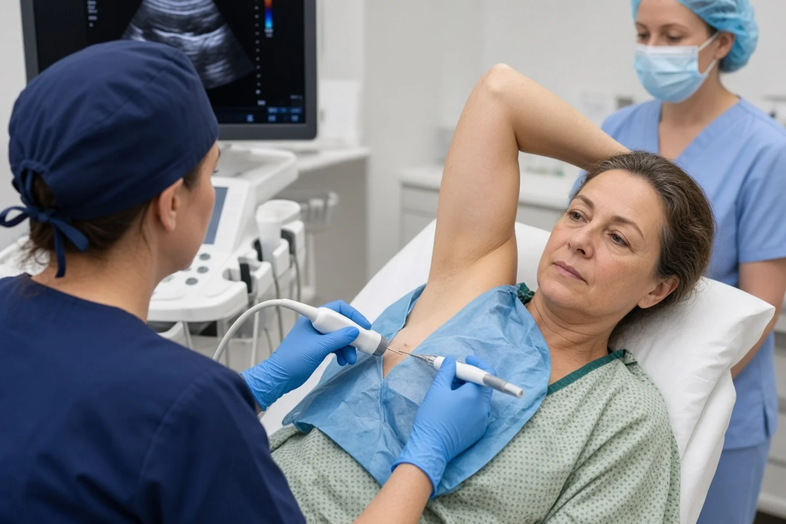
A new core needle biopsy, under local anaesthetic, confirms the diagnosis. The radiologist guides it by ultrasound, by mammography or, more rarely, by MRI, depending on how the area is best seen.
The pathology report says whether the lesion is a carcinoma in situ or an invasive cancer, its grade, and the markers tested by immunohistochemistry, a staining of the cells under the microscope: oestrogen receptors, progesterone receptors, HER2 protein. These markers may be different from those of the first cancer. The old analysis is therefore not enough on its own.
The time to result depends on the laboratory. A quick result is not, in itself, a bad sign. If a lymph node looks suspicious, it is also sampled, most often under ultrasound.
The staging work-up
When the recurrence is invasive, the guidelines generally provide for a staging work-up, to look for possible involvement of other organs before deciding on the operation. The 18-FDG PET scan is the examination most often offered.
If it is not available, a CT scan of the chest, abdomen and pelvis combined with a bone scan replaces it. In case of invasive lobular carcinoma, these two strategies can be used, because the PET scan is sometimes less informative.
For a purely in situ recurrence, the PET scan is not recommended. For a very small invasive focus confined to the breast, a staging work-up is not always necessary. Tumour markers do not make the diagnosis, but they can help in case of a rise over several successive blood tests.
The discussion at the multidisciplinary team meeting

Every recurrence file is discussed at a multidisciplinary team meeting before treatment. The surgeon, the oncologist, the radiation oncologist, the radiologist and the pathologist cross-check the imaging, the biopsy, the treatments already received and the staging work-up.
This meeting answers concrete questions: is it an isolated recurrence? Is a mastectomy the right procedure, or can a second breast-conserving treatment be discussed? Which procedure on the lymph nodes?
Is reconstruction possible at the same time? Is systemic treatment needed before or after the operation? Does further radiotherapy have a role?
You do not attend this meeting. Its conclusions are explained to you at the consultation, so that the decision is made with you.
A second opinion is always possible: bring the biopsy report, the images and, if there is one, the report of the multidisciplinary team meeting. See also how to choose a breast surgeon in Paris and prepare your first consultation.
The mastectomy, and the timing of the reconstruction, are prepared now.
Dr Zeitoun examines the breast and the armpit, reviews the imaging with you and explains the surgical options, including immediate or delayed reconstruction. Consultations at the practice at 241 rue du Faubourg Saint-Honoré (Paris 8th) and at Clinique Hartmann (Neuilly-sur-Seine). You can book an appointment on Doctolib, request a teleconsultation, or request a callback from the secretariat.
Surgery: often a mastectomy
When the staging work-up shows no distant disease, local treatment relies first on surgery.
After breast-conserving treatment and radiotherapy

If the first treatment was a lumpectomy — sometimes with oncoplastic surgery, which removes the tumour and reshapes the breast at the same time — followed by radiotherapy, the guidelines retain mastectomy as the recommended treatment of local recurrence. The breast has already received radiotherapy: a further breast-conserving operation, with no possibility of re-irradiation, is not recommended.
A second breast-conserving treatment is not the recommended treatment. It is rarely performed after breast cancer, but it is possible case by case, according to the precise indications of the guidelines, discussed at the multidisciplinary team meeting and with you. Mastectomy remains the recommended treatment.
The guidelines specify that this option can be discussed when the following conditions are all met at the same time: age over 60; a tumour expressing oestrogen and progesterone receptors, with a low Ki67, that is, a low proliferation marker, without excess HER2 protein, and without lymph node involvement, or a ductal carcinoma in situ of any grade; size of 2 cm or less; recurrence occurring more than five years after the first treatment; a unifocal lesion, that is, confined to a single focus; breast-conserving surgery feasible in good conditions, in particular for the cosmetic result and the condition of the skin after radiotherapy; information on the risks and the patient’s agreement. Partial breast irradiation is then discussed, in the absence of severe sequelae from the first radiotherapy. This option is not retained in the case of a lobular type, an invasive cancer associated with extensive ductal carcinoma in situ, or if there is a genetic predisposition to breast cancer.
The indication also takes into account the radiotherapy doses already received, the timing of the recurrence, the characteristics of the tumour and the toxicities of the first treatment. In these situations, a radiotherapy consultation takes place before the operation, to confirm the possibility of further irradiation and to assess the toxicities, in particular to the skin, of the first treatment.
If mastectomy is refused, breast-conserving surgery combined with accelerated partial breast irradiation can be an option, provided that the recurrence occurred more than five years after the first treatment.
This option carries a higher risk of a new local recurrence. It does not change overall survival. It also exposes to complications, in particular cosmetic ones, in a breast that has already been irradiated.
It is never an obligation. Mastectomy remains the recommended treatment.
When the lesion is not palpable, preoperative localisation (guide wire or magnetic clip) is organised by the radiologist the day before or the morning of the operation. The choice between keeping the breast or removing it, when it arises at the first treatment, is explained on the page lumpectomy or mastectomy. Here, the context is different: the breast has already been operated on and irradiated.
After a first mastectomy
When the recurrence is located on the chest after a mastectomy, the procedure most often consists of removing the affected area, with a margin of healthy tissue, once the staging work-up has been done. If there was a flat closure and there is a recurrence on the chest, no immediate reconstruction is done at that time, because the skin — or rather the skin envelope — has to be reconstructed.
Secondary reconstruction is done at a distance from any recurrence. What follows — radiotherapy, systemic treatment — depends on the extent of the lesion, the treatments already received and the discussion at the multidisciplinary team meeting.
The lymph nodes of the armpit
The procedure on the lymph nodes depends on what has already been done, and on the current imaging of the armpit.
If the first treatment included a sentinel lymph node procedure, the first lymph node relay for the lymph of the breast, and there are no signs of lymph node involvement, a new sentinel lymph node procedure can be offered in case of recurrence after breast-conserving treatment. After a mastectomy, no new sentinel lymph node procedure is done, because the mammary gland is no longer present. In this situation, a lymph node procedure, or even an axillary dissection, is discussed.
If an axillary dissection, the removal of several lymph nodes from the armpit, has already been done, a new procedure is not systematic. It is discussed if it would change the systemic treatment or the radiotherapy, or if a lymph node appears suspicious and the biopsy confirms it. If a lymph node is already known to be involved before the operation, the procedure is adapted at the multidisciplinary team meeting.
The aftermath of a procedure on the armpit, and the useful steps to limit swelling of the arm, are described on the page lymphoedema prevention.
Reconstruction, immediate or delayed
Reconstruction is not a treatment for the cancer, and it is not compulsory. You may wish to regain breast volume, or a flat closure. This decision can also be made later.
After breast cancer irradiated following breast-conserving treatment, if a mastectomy is performed and reconstruction is wished for, immediate reconstruction with a flap is readily offered. Reconstruction with an implant is possible, but often advised against in view of the risks and postoperative complications.
If there was a flat closure and there is a recurrence on the chest, no immediate reconstruction is done at that time, because the skin — or rather the skin envelope — has to be reconstructed. Secondary reconstruction is done at a distance from any recurrence.
The options usually discussed are reconstruction with an implant, sometimes in several stages; a latissimus dorsi flap, which uses the muscle and skin of the back; a DIEP flap, which uses the skin and fat of the abdomen, performed with a microsurgery team; a gracilis flap, taken from the inner thigh, also with a microsurgery team; lipofilling, which reinjects your own fat, most often as a complement once the tissues have stabilised; then, once the volume has been restored, nipple reconstruction and areola tattooing. After previous radiotherapy, the flap is the technique readily offered for immediate reconstruction.
Preserving the skin or the nipple-areola complex (NAC) at the time of the mastectomy is explained on the page dedicated to preserving the nipple-areola complex.
Several operations, spread over 18 to 24 months, are sometimes necessary for touch-ups. The result can be very beautiful, while remaining different from the breast before. The page breast reconstruction presents all the techniques.
Dr Zeitoun practises in Sector 2 (non-OPTAM) and charges fees above the French Social Security rate for all consultations and procedures. Most international patients are not covered by French Social Security. A detailed quote is given during the consultation, before any procedure. If you are covered, the Assurance Maladie reimburses on the basis of the Social Security rate, and your health insurance may cover all or part of the additional fees, depending on your policy.
Practical advice before the operation is on the page preparing for your operation, and advice for the first weeks on the page postoperative recovery.
Associated treatments
Breast surgery is only one part of the treatment. Systemic treatment — chemotherapy, hormone therapy, immunotherapy or targeted therapy — and, sometimes, further radiotherapy are discussed in all cases at the multidisciplinary team meeting.
Systemic treatment relies on the new biopsy, not only on the old one: hormone receptors, HER2, in situ or invasive nature, size, lymph nodes, time since the first cancer, and treatments already received. The genomic tests used for some first hormone-sensitive cancers are not validated in case of local recurrence.
Depending on the situation, the following may be discussed: hormone therapy if the recurrence has hormone receptors; chemotherapy, especially in case of an invasive recurrence, an aggressive form or a short interval since the first treatment; targeted therapy, for example an anti-HER2 treatment if the HER2 protein is in excess; immunotherapy, mainly discussed in certain triple-negative forms.
Systemic treatment before the operation, called neoadjuvant chemotherapy when it is chemotherapy, is not the standard when a local recurrence, in an area already irradiated, can be operated on straight away. It can however be discussed in aggressive forms, in particular HER2-positive or triple-negative, adapting it to the treatments already received.
If the breast or the chest has not yet been irradiated, radiotherapy is often discussed after the operation. If radiotherapy has already been given, partial re-irradiation can be considered as part of a second breast-conserving treatment, after the radiation oncologist’s opinion. Referral for a radiotherapy consultation is discussed case by case.
When the first cancer did not lead to genetic testing, it can be offered according to the same criteria as at diagnosis: age, triple-negative form, involvement of both breasts, personal or family history. A mutation of the BRCA1 or BRCA2 genes, or of PALB2, can then open a discussion on a preventive mastectomy of the other breast and, if necessary, on preventive removal of the ovaries and fallopian tubes.
It is never an obligation, and the decision can be made at a distance from the treatments. See also positive BRCA result and hereditary breast and ovarian cancer risk.
If the staging work-up shows distant disease, the treatment becomes mainly systemic. Breast surgery then has only a limited place, discussed case by case.
What can be said about the prognosis, without false precision
An isolated local recurrence is not, in itself, the announcement of a generalised disease. When it is confined to the breast or the chest, after a reassuring staging work-up, treatment is undertaken with the aim of cure.
Several factors weigh more than a single figure, which no one can invent for your situation: the type of the recurrence, a carcinoma in situ not having the same significance as an invasive cancer; the time since the first treatment, a recurrence occurring after several years not having the same meaning as an early recurrence; the size, the grade, lymph node involvement; the markers (hormone receptors, HER2); the absence or presence of distant lesions.
The subtype of the first cancer also matters. A recurrence of a hormone-sensitive cancer, a HER2-positive cancer or a triple-negative cancer is not discussed in the same way.
Age at the time of the recurrence, and the treatments already received, enter into the same reflection. The prognosis is discussed with you, on the basis of your file, not on the basis of a percentage read out of context.
Follow-up after treatment of a recurrence
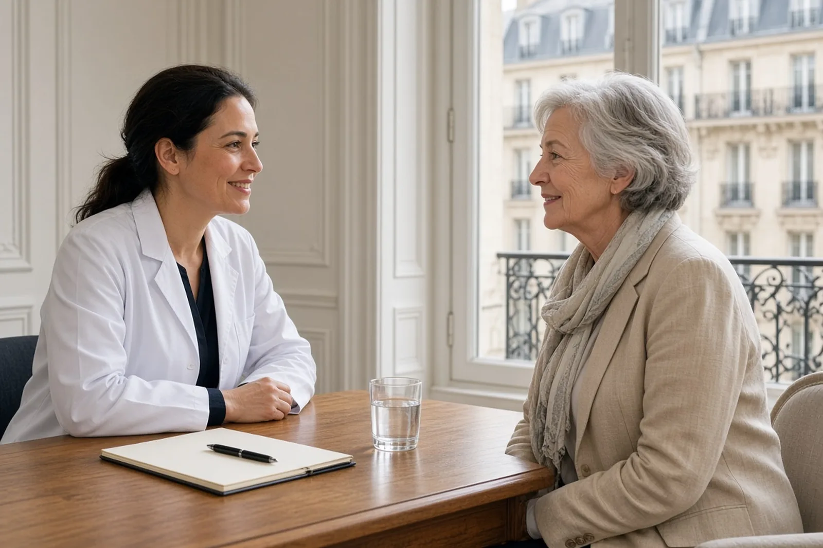
Once the treatments are over, follow-up resumes on a regular and prolonged basis. After breast cancer, the guidelines provide for a clinical examination every six months for five years, then once a year, and a mammogram and an ultrasound every year, for life.
The first mammogram is done a few months after the end of any radiotherapy. This schedule is adapted to your situation.
After a mastectomy, surveillance of the operated side is clinical: there is no longer a breast to X-ray on that side. The other breast, if it is still there, continues to be followed with a yearly mammogram and ultrasound, and sometimes by MRI depending on the context.
Follow-up can be shared in alternation between the surgeon, the oncologist, the radiation oncologist and the gynaecologist. No other whole-body imaging examination is systematic in the absence of symptoms.
Report without waiting for the next visit a new lump, a change in the scar, a lymph node, bone pain that persists or an unusual cough. These signs do not mean that the disease has come back; they simply need to be examined.
Do questions remain, or would you like your file reviewed?
Dr Zeitoun can go over the work-up, the surgical options and the follow-up schedule with you. Consultations at the practice at 241 rue du Faubourg Saint-Honoré (Paris 8th) and at Clinique Hartmann (Neuilly-sur-Seine). Teleconsultation, appointment on Doctolib, or request a callback from the secretariat.
Frequently asked questions
The questions asked most often after the announcement of a local recurrence. If yours is not here, ask it at the consultation.
Does a local recurrence mean that the cancer has spread throughout the body?
No. A local recurrence is located in the breast that has already been treated or on the chest. Distant disease, in another organ, is called a metastasis. It is the role of the staging work-up to check this distinction before deciding on the operation.
Is a mastectomy always necessary?
After breast-conserving treatment and radiotherapy, mastectomy is the recommended treatment. A second breast-conserving treatment is not the recommended treatment. It is rarely performed, but it can be discussed case by case, according to the precise indications of the guidelines, at the multidisciplinary team meeting. A radiotherapy consultation is then discussed case by case, to examine the possibility of further irradiation. It is not recommended to repeat a lumpectomy without the possibility of further irradiation.
Can the breast be reconstructed straight away, even though it has already been irradiated?
After breast cancer irradiated following breast-conserving treatment, if a mastectomy is performed and reconstruction is wished for, immediate reconstruction with a flap is readily offered. An implant is possible, but often advised against in view of the risks and postoperative complications. If there was a flat closure and there is a recurrence on the chest, no immediate reconstruction is done at that time, because the skin — or rather the skin envelope — has to be reconstructed. Secondary reconstruction is done at a distance from any recurrence. The decision is made with you, taking into account the condition of the skin, your body shape and what you wish.
Does the recurrence necessarily have the same type as the first cancer?
Not necessarily. Hormone receptors and HER2 status can change. That is why a new biopsy is essential. The pathology report of this biopsy guides the associated treatments.
How is the staging work-up done?
For an invasive recurrence, a PET scan is most often offered. If it is not available, a CT scan of the chest, abdomen and pelvis is combined with a bone scan. For a purely in situ recurrence, the PET scan is not recommended. Tumour markers do not make the diagnosis, but they can help in case of a rise over several successive blood tests.
What is the prognosis of a local recurrence?
It depends on the type (in situ or invasive), the time since the first treatment, the lymph nodes, the markers and the absence of distant lesions. An isolated local recurrence is treated with the aim of cure. No single percentage applies to all situations.
Should I have a genetic test?
If testing was not done at the time of the first cancer, it can be offered according to the guidelines: age, triple-negative form, personal or family history, involvement of both breasts. The result also concerns your relatives, if they wish to be informed and, where appropriate, tested. See the page hereditary breast and ovarian cancer risk.
Is pain near the scar a sign of recurrence?
After a lumpectomy or a mastectomy, pain related to the small nerves of the chest is common. A change in the scar, a new lump or a change in the skin should be examined, with imaging and, if needed, a biopsy. See also pain after a mastectomy.
How does surveillance work afterwards?
A clinical examination every six months for five years, then once a year, and a yearly mammogram and ultrasound of the remaining breast and of the other breast, for life. After a mastectomy, the operated side is followed by clinical examination. Report any new sign without waiting for the next visit.
Can I ask for a second opinion?
Yes, it is your right, and it does not delay treatment when it is organised quickly. Bring the biopsy report, the images, the reports of the treatments already received and, if there is one, the report of the multidisciplinary team meeting. See second opinion for breast cancer.
What are Dr Zeitoun’s fees?
Dr Zeitoun practises in Sector 2 (non-OPTAM) and charges fees above the French Social Security rate for all consultations and procedures. Most international patients are not covered by French Social Security. A detailed quote is given during the consultation, before any procedure. If you are covered, the Assurance Maladie reimburses on the basis of the Social Security rate, and your health insurance may cover all or part of the additional fees, depending on your policy.
Sources
This article is based in particular on the SENORIF 2025-2026 guidelines.
- SENORIF 2025-2026 guidelines — Breast cancers and diseases: diagnostic and therapeutic approaches, treatment protocols (in French): staging work-up in case of suspected recurrence, management of local recurrences, surgery, associated treatments, follow-up. Public page of the guidelines: https://curie.fr/senorif-le-referentiel-de-traitement-des-cancers-et-pathologies-du-sein
- French National Authority for Health (HAS) — Breast reconstruction or flat closure: your wishes and options, shared decision aid, 29 March 2023 (in French). https://www.has-sante.fr/jcms/p_3351381/fr/reconstruction-mammaire-ou-buste-plat-vos-souhaits-et-options
- French National Authority for Health (HAS) — Autologous breast reconstruction techniques, alternatives to breast implants (in French). https://www.has-sante.fr/jcms/c_2965016/fr/techniques-de-reconstruction-mammaire-autologues-alternatives-aux-implants-mammaires
- Loibl S. et al. — Early breast cancer: ESMO Clinical Practice Guideline for diagnosis, treatment and follow-up. Annals of Oncology, 2024. https://www.annalsofoncology.org/article/S0923-7534(23)05104-9/fulltext
- American guidelines — NCCN Guidelines for Patients: Invasive Breast Cancer. https://www.nccn.org/patients/guidelines/content/PDF/breast-invasive-patient.pdf