You have just been offered a mastectomy for a breast cancer. A mastectomy removes the breast gland. Can the nipple and areola be preserved?
The areola is the more deeply pigmented skin around the nipple. Together they form the nipple-areola complex (NAC). The skin envelope is the skin that covers the breast. The operation that preserves this envelope and the NAC is called a nipple-sparing mastectomy. The operation as a whole is described on the mastectomy page. The choice of when to reconstruct is described on the page about immediate or delayed reconstruction.
This article is based in particular on the SENORIF 2025-2026 guidelines.
Dr Zeitoun examines the breast and reviews your images with you to see whether the nipple and areola can be preserved.
REQUEST A CALLBACKBOOK ON DOCTOLIB →
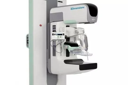
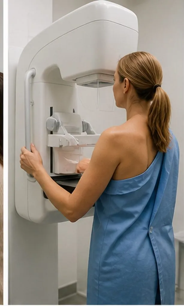
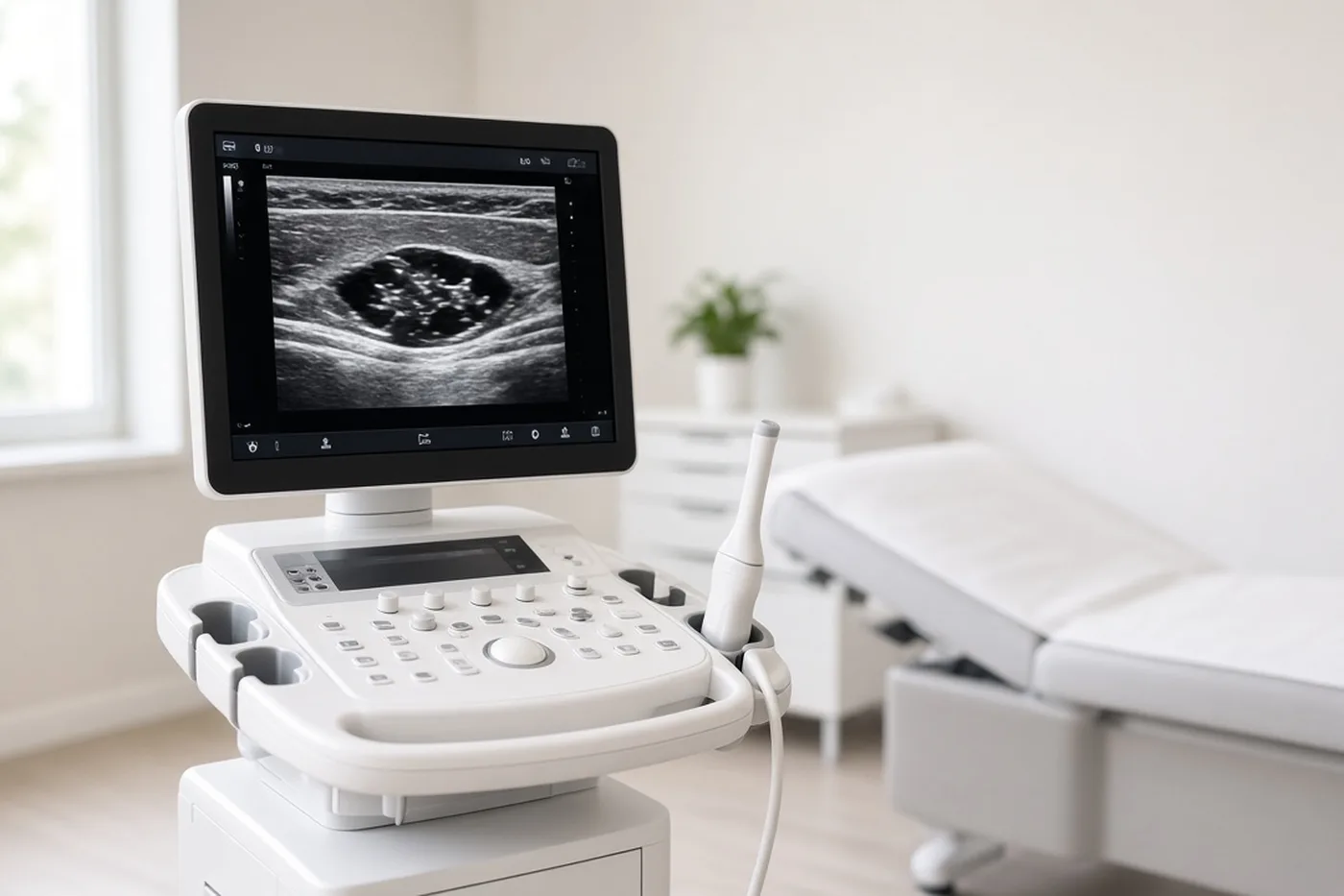
Regarding breast cancer, the nipple and areola are preserved when imaging done before the operation places the lesion more than 1 cm from the NAC. This distance is read on the mammogram, on the ultrasound and, when it has been done, on the breast MRI (magnetic resonance imaging). If there are several images, the one closest to the NAC is the one that counts. If the distance between the lesion and the NAC is less than 1 cm, the NAC must be sacrificed.
This distance between the lesion and the NAC must appear in the imaging report.
The second constraint that can require removal of the NAC depends on cup size and ptosis ("drooping breasts"). The greater the ptosis, the greater the risk of a poorer blood supply to the skin envelope, and therefore to the NAC. To avoid secondary necrosis, it may have to be sacrificed.

Preserving the NAC is recommended for a mastectomy that treats a breast cancer already known, when immediate reconstruction takes place during the same operation. Besides the distance, three conditions are met.
If one of these three points is present, the NAC is not preserved, even if the main lesion looks far away. Discharge is investigated before any decision is made.
A preserved NAC is fed by the small vessels of the skin envelope. The larger the cup size and the greater the ptosis, the less reliable these vessels are, and the higher the risk of secondary necrosis. Smoking, diabetes and previous radiotherapy to the chest also weaken this blood supply.
Each situation is assessed individually, in consultation.
When the mastectomy involves both breasts, each breast is assessed separately: the NAC may be preserved on one side and sacrificed on the other.

When the NAC is preserved, the surgeon removes a retroareolar specimen (the tissue located just behind the areola) during the operation and sends it for analysis, so as not to miss a cancer located behind the NAC.
It is not recommended to perform a routine frozen section. A frozen section is a reading under the microscope while you are asleep. The answer for the retroareolar tissue does not come during surgery, but rather 10 to 15 days later, at your postoperative consultation, where you are given the histology results.
If this specimen contains an invasive (spread beyond the ducts) or in situ (still confined to the ducts) breast cancer, and the margins, meaning the distance between the abnormal cells and the edge of the removed tissue, are not enough under the definitions in the recommendations, a secondary removal of the NAC may be offered during a second operation. Atypia alone, meaning unusual cells that are not a breast cancer, does not lead to that second operation.
The nerves that run through the gland are cut with it. After a mastectomy, overall sensation in the breast is lost, whether the nipple is preserved or not. A microscopic nerve repair, called neurotization, aims to bring back part of the sensation. It is a very highly specialised technique, offered in very few centres. It is not the usual operation here, and no result is promised.
Colour can change. So can projection: the nipple can flatten. The areola can become paler, darker, or no longer match the other side.
Necrosis is the main risk when skin and NAC are preserved. In the days that follow, the nipple turns dusky, then black over part of the NAC or over all of it. Limited necrosis heals and leaves a crust and a loss of colour. Extensive necrosis means the nipple is lost. A second operation is sometimes needed. Later, if you wish, projection and colour can be rebuilt. That stage is on the page about areola tattoo. It is not decided on the day of the mastectomy.
Stopping smoking before the operation protects these small vessels.
If you choose flat closure, meaning without recreating the volume of the breast, the NAC is not preserved.
Immediate reconstruction can be offered with a mastectomy for a breast cancer. Immediate means under the same general anaesthetic as the mastectomy. You wake with a volume in place. Delayed means later. The choice of timing is on the page about immediate or delayed reconstruction. Preserving the nipple is not a condition for reconstruction: reconstruction is also done when the NAC has been removed. Skin that is preserved, with or without the nipple, can stay only if the volume is rebuilt during the operation. Once the skin has closed flat, that envelope is not recovered.
An implant, a flap from the abdomen (DIEP, which uses skin and fat from the lower abdomen), a latissimus dorsi flap, or fat transfer: a flap is tissue taken from elsewhere on the body. A nipple that is preserved changes the envelope that is available. It does not choose the technique.
Radiotherapy, treatment with rays, can be indicated after mastectomy according to the tumour. This indication stays the same whether or not the NAC was preserved. Preserving the NAC must not delay the start of the treatments that follow the operation. Previous radiotherapy to the chest, on the other hand, weighs on the risk of necrosis. If radiotherapy is planned after the operation, the reconstruction technique is discussed one case at a time: an implant may be chosen, because it can be changed later, whereas an irradiated flap preserves the effects of the rays. The detail is on the immediate reconstruction page.
Inflammatory breast cancer, a rare form of breast cancer in which the breast becomes red, hot and swollen, rules out reconstructing during the same operation. In this form, the nipple question does not arise in the same way, because the volume is not rebuilt during the operation.
Bring your imaging and the report to Dr Zeitoun: it must state the distance between the lesion and the nipple-areola complex.
REQUEST A CALLBACKBOOK ON DOCTOLIB →
The incision is chosen according to the shape of the breast, the skin, and the technique used for volume. When the NAC stays, three sites are used, described on the mastectomy page: the fold under the breast, the armpit, and the edge of the areola. The fold under the breast is the one chosen most often. These scars sit in a fold or at an edge, so they show little in a neckline. When the NAC is removed and the skin stays, the scar is oblique, at the centre of the breast.
A route with a camera, called endoscopic, under the breast or under the arm, or a robot-assisted route is discussed only if the nipple stays. They are for nipple conservation with immediate reconstruction by implant. They are not available everywhere. The consultation says whether they have a place in your case.

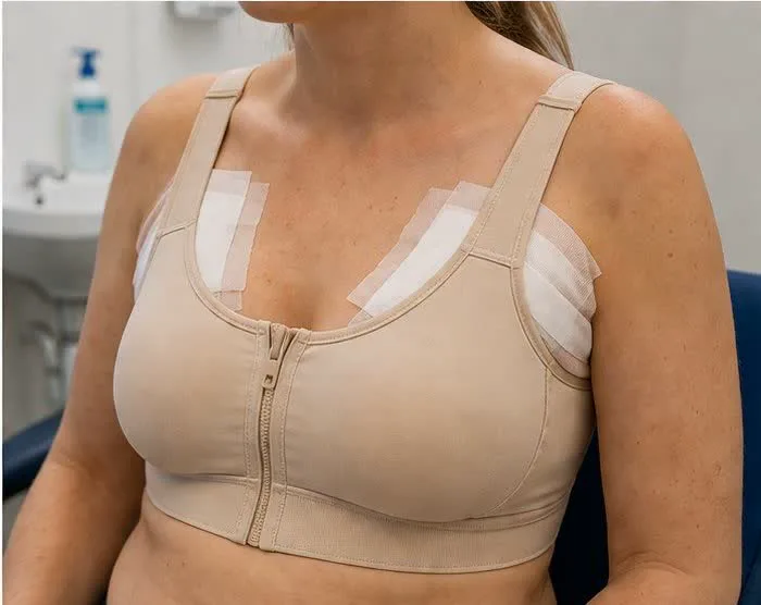
In the first days, the colour of the nipple is watched. It is the sign that blood is arriving. An area that darkens is examined without waiting for the next appointment. The timetable for drains, the support bra, and the return to activity is on the mastectomy page.
Breastfeeding is no longer possible from the breast whose gland has been removed. A nipple that is preserved does not replace it.
There is no longer a gland to read with a mammogram on the operated side. Follow-up rests on examination of the skin, the scar, and the nipple. An ultrasound or an MRI is added according to your situation. A new crust, discharge, or a lump under the areola is shown to the surgeon.

The answer about the nipple and areola is not given by email, or from a photograph alone. It requires the images (mammogram, ultrasound, MRI if there is one) and examination.
The surgeon looks at how far the breast droops, the skin, smoking, diabetes, and any previous radiotherapy. He says what is realistic for the NAC, and what belongs to volume, on the mastectomy page as on the immediate reconstruction page. He explains necrosis, a change in colour, loss of projection, loss of sensation, and a possible secondary removal if the analysis behind the areola is abnormal.
Before surgery for a breast cancer, it is recommended to hold a multidisciplinary meeting. Several doctors then review the records together. That meeting does not replace examination of the nipple.
Consultations at the practice, 241 rue du Faubourg Saint-Honoré (Paris 8th), and at Clinique Hartmann (Neuilly-sur-Seine). Surgery takes place at Clinique Hartmann.
Sector 2, not opted into OPTAM, with possible fee supplements. The amount is explained during the consultation and written on a quote before the procedure.
Yes, when imaging before the operation places the lesion more than 1 cm from the NAC, and the other conditions in the recommendations are met. If the distance is less than 1 cm, the NAC must be sacrificed. Cup size, ptosis and the risk of secondary necrosis can also require its removal. The operation itself is described on the mastectomy page.
The mammogram, the ultrasound and, when it has been done, the MRI. If there are several lesions, the one closest to the NAC decides.
The NAC must be sacrificed. The skin of the breast can stay. The scar is then oblique, at the centre. Projection and colour can be rebuilt later, if you wish, as described on the page about areola tattoo.
Yes. It is recommended to preserve the NAC only in the absence of discharge, of Paget's disease and of microcalcifications within one centimetre of the nipple.
Not automatically. The greater the ptosis, the greater the risk of a poorer blood supply to the skin envelope, and therefore to the NAC. To avoid secondary necrosis, the NAC may have to be sacrificed. Each situation is assessed individually.
No. After a mastectomy, overall sensation in the breast is lost, whether the nipple is preserved or not. Neurotization, a very highly specialised technique offered in very few centres, is not the usual operation and promises no result.
Yes. That is necrosis. Part of the NAC, or all of it, can be lost. Colour is watched from the first days. Smoking, diabetes, thin skin or previous radiotherapy to the chest make this risk more concrete.
If the retroareolar specimen contains a cancer and the margins are not enough, a secondary removal of the NAC may be offered during a second operation. Atypia alone does not lead to that removal. There is no routine frozen section.
Yes. That is the setting in which the recommendations describe preserving the NAC: during immediate reconstruction. Implant, flap, or fat transfer are chosen separately. Preserving the nipple is not a condition for reconstruction. If you choose flat closure, the nipple is not preserved.
The indication for radiotherapy after the operation is the same whether or not the NAC was preserved. Previous radiotherapy to the chest, on the other hand, can increase necrosis. If radiotherapy is planned after mastectomy, the technique used for volume is discussed on the page about immediate or delayed reconstruction. Preserving the NAC must not delay the treatments that follow.
Yes. Inflammatory breast cancer, a red, hot and swollen breast, rules out reconstructing during the same operation. Preserving the nipple is discussed together with a volume rebuilt during the operation. In this form, that condition is not met.
Not from the breast whose gland has been removed. A nipple that is preserved does not produce milk.
Further reading
Mastectomy in Paris: the surgeon removes the breast for cancer. Skin or nipple sometimes spared. Immediate or delayed reconstruction. Paris 8th, Hartmann.
ReconstructionBreast reconstruction at the time of mastectomy or later: what guides the timing, including radiotherapy. Dr Zeitoun, Paris.
ReconstructionBreast implant reconstruction, immediate or delayed, with or without an expander. Consultations in Paris 8th and at Clinique Hartmann; surgery at Hartmann.
Dr Zeitoun sees you at the Faubourg Saint-Honoré practice or at Clinique Hartmann to go through the file with you.
REQUEST A CALLBACKBOOK ON DOCTOLIB →