


Each armpit contains dozens of lymph nodes. They are small filters of the immune system, draining the arm, the chest wall and the breast. At rest they measure a few millimetres and cannot be felt. When they are working, they swell: that is their job.
A lump in the armpit is therefore not, in itself, a sign of cancer. Most often it reflects a passing reaction: a small cut on the hand, folliculitis after shaving, a cold, or a vaccine given in that arm. Sometimes it is not even a lymph node, but accessory breast tissue, a skin cyst or a lipoma.
A few signs, on the other hand, call for a check-up without delay. Knowing them helps avoid two pitfalls: panicking over a reactive node, or leaving a lump that does not go down.
A lump under the arm, an ultrasound that mentions a “lymph node”: should you worry?
Dr Jérémie Zeitoun, surgical oncologist and breast surgeon, sees patients at the practice at 241 rue du Faubourg Saint-Honoré (Paris 8th) and at Clinique Hartmann (Neuilly-sur-Seine). Bring the reports and images you already have. Surgery, when it is useful, takes place at Hartmann. Consultations can be held in English.
A swollen lymph node: most of the time, it is doing its job

The lymphatic system is a network of small vessels that carries fluid from the tissues back into the circulation. Along the way, lymph nodes trap germs and organise the immune defence. When an infection, even a minor one, affects the hand, the arm or the skin of the armpit, the nodes draining that area enlarge for a few days or weeks.
A “reactive” node is often soft, mobile under the finger, sometimes tender. It appears together with a visible cause, then shrinks once that cause has cleared.
By contrast, a node that is hard, fixed to the surrounding tissue, painless, growing, or that persists with no explanation deserves a medical opinion. These are not proof of anything serious; they are reasons to take a closer look.
Other lumps can form in the armpit
An epidermoid cyst (so-called sebaceous cyst), a lipoma (a small, soft fatty lump), a boil or a flare of hidradenitis suppurativa, or accessory breast tissue, which swells before periods and during breastfeeding. Clinical examination and ultrasound tell them apart within minutes.
Infection, vaccine, skin: the usual explanations

What makes an armpit node swell
- A minor infection of the arm or hand. A cut, an insect bite, a whitlow, an ingrown nail, infected eczema. The entry point is sometimes forgotten.
- The skin of the armpit. Folliculitis after shaving or waxing, a boil, hidradenitis suppurativa.
- A Covid-19 vaccine given in that arm. Within 12 weeks of the injection, it often makes the nodes on the injection side react.
- A cat scratch. “Cat scratch disease” causes a node that is often slightly painful and may last several weeks. Your GP sometimes prescribes an antibiotic.
- A general infection. Glandular fever, toxoplasmosis, a cold or a sore throat: several groups of nodes then react at the same time, often with fever and tiredness.
- A breast implant. If silicone leaks, a lymph node can take it up and enlarge. Ultrasound recognises it; the implant reconstruction page describes how the implant is monitored.
Noting when the lump appeared, its approximate size and any possible cause (vaccine, cut, fever) helps the doctor a great deal later on.
Causes that need investigating
An armpit node can also reveal breast cancer (in women as in men), lymphoma or, more rarely, another tumour such as a melanoma of the arm or back. This is rare compared with the number of reactive nodes, but it is exactly why a node with no explanation should be examined.
When to see a doctor, and how soon
See a doctor without waiting
A node together with a change in the breast on the same side: a lump, skin or nipple retraction, red and thickened skin, nipple discharge. The breast and armpit are then assessed together.
See a doctor promptly
No obvious cause; a node that is hard, fixed or growing; persistent fever, night sweats, weight loss; a history of breast cancer, melanoma or lymphoma.
Keep an eye on it
A soft, mobile, slightly tender node with an obvious cause: a recent vaccine in that arm, a small cut, folliculitis, a flu-like illness. It is reasonable to keep an eye on it. If it does not go down, or if it grows, mention it to your doctor.

What the doctor checks
The doctor asks when the lump appeared, about vaccines, cuts, fever, treatments and past history. They examine both breasts, both armpits, the hollow above the collarbone, the neck, and the skin of the arm and back. A single node does not mean the same as several nodes in different areas.
Your GP or gynaecologist may, depending on the situation, order a blood test and an ultrasound. A breast specialist consultation becomes useful when the ultrasound describes an “atypical” or “suspicious” node, or when there is a change in the breast.
Ultrasound first, mammogram depending on age and context
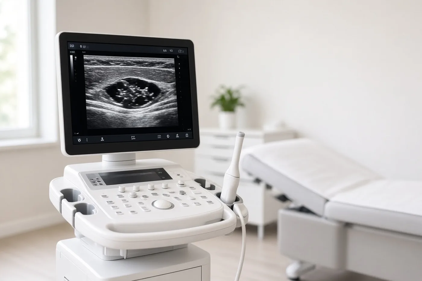
An ultrasound of the armpit is the key test. It is painless, uses no radiation and takes a few minutes. It first shows whether the lump really is a lymph node, then describes how it looks.
What the radiologist looks at
- A normal-looking node is oval, with a clearly visible fatty centre (the “hilum”) and a thin outer layer (the “cortex”). Even if slightly enlarged, this appearance is reassuring.
- A node to monitor or sample is rounder, with a thickened cortex, a fading hilum, or unusual blood flow on Doppler.
- The breasts are examined at the same time. In women, a mammogram is usually added, in particular if the node looks suspicious.
The report ends with an ACR (BI-RADS) category. A typical reactive node on the side of a recent vaccine, with no breast abnormality and no history of cancer, is classified as benign: according to the European breast imaging recommendations (EUSOBI), no further test is needed. Professional societies also stress one point: a vaccine should not delay a mammogram. Simply tell the radiologist the date and the arm of the injection.
The images, not just the text
Recent images and reports, plus previous scans for comparison. The preparing your first consultation page lists what is useful. A breast MRI is not a first-line test for a lymph node: it is discussed in specific situations, described below.
The ultrasound describes an “atypical” node or suggests a biopsy.
Dr Zeitoun reviews the images and report with you, examines the breasts and armpits, and tells you whether to monitor, sample or operate. Consultation at the practice at 241 rue du Faubourg Saint-Honoré (Paris 8th) and at Clinique Hartmann (Neuilly-sur-Seine); surgery at Hartmann.
When the node is sampled: what the result changes

If the node does not look typically benign, the radiologist suggests an ultrasound-guided core biopsy. It is done under local anaesthetic in a few minutes, much like a breast biopsy. Several small samples are taken and sent to the pathology laboratory. A simple fine-needle aspiration is sometimes done instead.
The result usually comes back within a few days to one or two weeks; the biopsy results timing page explains what makes this vary. A small marker clip is often placed in the node during the biopsy, so it can be found later.
Reactive, benign node
The tissue is inflamed, with no abnormal cells, and the result matches the images. The node is monitored, usually with a clinical examination and a follow-up ultrasound after a few months.
Inconclusive sample
The samples are too small, or the result does not match the images. A repeat biopsy, or surgical removal of the node, is then discussed.
The breast work-up takes centre stage
If the node contains cells coming from the breast, the work-up looks for the tumour in the breast: mammogram, ultrasound, then breast MRI if those are normal. The case is discussed at a multidisciplinary team meeting, and treatment follows the breast cancer pathway, sometimes with chemotherapy before surgery.
Another specialist takes over
Lymphoma is managed by a haematologist. To classify it precisely, the whole node often needs to be analysed: surgical removal is then requested. Another origin (skin, melanoma…) leads to the relevant specialist.
When the surgeon steps in

The vast majority of armpit nodes are never operated on. The surgeon steps in for specific situations.
Surgical situations
- Removing a node to analyse it whole. When the needle biopsy was inconclusive, or when the haematologist needs the complete node. It is a short operation, usually as a day case, through an incision of a few centimetres hidden in a skin fold of the armpit.
- A node that cannot be felt is removed after preoperative localisation (wire or magnetic clip), placed by the radiologist, to go straight to the right target.
- A lump that is not a lymph node and is bothersome: cyst, lipoma, accessory breast tissue. Removal is discussed according to the discomfort, not as a matter of principle.
- Breast cancer with lymph node involvement. Armpit surgery is then part of the treatment: sentinel node biopsy or axillary dissection, decided at a multidisciplinary meeting, often after chemotherapy.

After a node has been removed
You can move your arm the same evening. The scar, in the skin fold, is usually very discreet. Removing a single node rarely causes arm swelling; useful measures are described on the lymphoedema page. The pathology result is given in consultation one to two weeks later, together with the next steps. Practical advice before the operation is on preparing for surgery.
Common questions
The questions that come up with a lump in the armpit. If yours is not here, it belongs in the consultation, or with Sophie, at the bottom right of the screen.
Is a lump in the armpit always cancer?
No. In the vast majority of cases it is a lymph node reacting to an infection, a vaccine or irritated skin, or another benign lump (cyst, lipoma, accessory breast tissue). A check-up is useful when the lump is hard, fixed, growing, persists with no cause, or comes with a change in the breast.
How long should I wait before seeing a doctor?
With an obvious cause (vaccine, cut, infection) and a soft, mobile node, keeping an eye on it is reasonable; if it does not go down, mention it to your doctor. With no cause, or if the node is hard, fixed or growing, or if there is a breast lump, lasting fever or weight loss, see a doctor without waiting.
Is a painful node more reassuring?
Often, yes: pain tends to go with inflammation or infection. But it is not an absolute rule. What counts is the whole picture (cause, consistency, how it changes, breast examination), and ultrasound settles any remaining doubt.
I was vaccinated recently. Should I postpone my mammogram?
No. Breast imaging recommendations are clear: vaccination should not delay a mammogram. Simply tell the radiologist the date and the arm of the injection. A node on the same side, with no other abnormality and no history of cancer, is considered benign.
Can deodorant or hair removal cause cancer?
No. No study has shown that deodorants or antiperspirants cause breast cancer. Shaving and waxing can, however, irritate the skin and make a node react for a few days.
Do I need a blood test?
Sometimes. Your GP orders one depending on the situation: blood count, inflammation markers, and certain serologies (glandular fever, toxoplasmosis, cat scratch disease). It complements the ultrasound; it does not replace it.
The ultrasound mentions an “atypical” node. What does that mean?
That its appearance is not typically reactive: thickened cortex, rounder shape, less visible fatty centre. It is not a diagnosis. Depending on the context, the radiologist suggests a check after a few weeks or an ultrasound-guided biopsy.
Does a lymph node biopsy hurt?
It is done under local anaesthetic and takes a few minutes. You mainly feel pressure and hear the device. A small bruise is common for a few days. The procedure is very similar to a breast biopsy.
The node contains cells from the breast, but the mammogram is normal. What happens next?
This is rare. A breast MRI is then performed, to look in the breast for a lesion that the mammogram and ultrasound did not show. The case is discussed at a multidisciplinary meeting, and treatment follows the breast cancer pathway.
Is removing a node the same as an axillary dissection?
No. Removing a single node to analyse it is a short, limited procedure. Sentinel node biopsy and axillary dissection are part of breast cancer treatment, decided at a multidisciplinary meeting.
Can men have an armpit node related to the breast?
Yes, although it is rare. The work-up is the same: examination, ultrasound, mammogram if needed. Male breast cancer has its own page.
I have had breast cancer before. Should a node worry me?
It should be examined without waiting several weeks. Tell the team that follows you. Many of these nodes remain reactive, but ultrasound, and a biopsy if needed, can confirm this quickly.
Sources
This article is based in particular on the SENORIF 2025-2026 guidelines.
- French SENORIF 2025-2026 reference — Breast cancers and breast disease: diagnostic and therapeutic approaches: work-up of a breast or armpit abnormality and multidisciplinary team discussion.
- Schiaffino S. et al. — European Society of Breast Imaging (EUSOBI) guidelines on the management of axillary lymphadenopathy after COVID-19 vaccination: 2023 revision. Insights into Imaging, 2023.
- Flegg et al. — Canadian Association of Radiologists and Breast Radiologists of Canada: axillary lymphadenopathy following vaccination, 2026 update. Canadian Association of Radiologists Journal, 2026 (doi 10.1177/08465371261450192).
- Society of Breast Imaging — recommendations for managing axillary adenopathy after vaccination, February 2022 revision: do not delay mammograms.
- American College of Radiology — BI-RADS Atlas: classification of breast and axillary imaging findings.
- French National Cancer Institute (INCa) — e-cancer.fr: breast cancer, symptoms and common misconceptions.
A node with no explanation, a result to understand.
Dr Jérémie Zeitoun sees patients at the practice at 241 rue du Faubourg Saint-Honoré (Paris 8th) and at Clinique Hartmann (Neuilly-sur-Seine) and operates at Hartmann. Bring the ultrasound, the mammogram and, if available, the biopsy report.