


This article draws in particular on the SENORIF 2025-2026 guidelines.

Has your biopsy report concluded that the lesion is benign or “at-risk”, and has an excision been proposed?
Come with this document and your imaging examinations: at the consultation, Dr Jérémie Zeitoun goes through your file with you and explains why the operation is proposed, or why surveillance may be considered.
A lumpectomy for a benign lesion: what does it mean?
A lumpectomy removes a limited area of the breast, the one that contains the lesion, and keeps the rest of the breast. It mainly concerns benign at-risk lesions. Benign lesions are monitored; a fibroadenoma, entirely benign, may nevertheless be removed in specific situations.
It is part of the surgery of benign breast lesions.
Removing the lesion to analyse it in full
The biopsy samples a fragment of the suspicious area, not the whole area. Analysis of the area removed makes it possible to be sure that there is no cancer next to it. The result is given in the pathology report.
What sets it apart from a lumpectomy for cancer
The procedure resembles a lumpectomy for cancer, but its rules are not the same. Breast cancer imposes requirements that do not apply to benign lesions:
- Margins. For an at-risk lesion such as atypical ductal hyperplasia, if the excision is not complete, a further operation is not necessary.
- Lymph nodes. For a benign lesion removed by lumpectomy, the lymph nodes of the armpit are not analysed: no sentinel lymph node.
- Radiation. After a lumpectomy for a benign lesion, there is no radiotherapy, except for pleomorphic lobular carcinoma in situ or lobular carcinoma in situ with necrosis, and for borderline or malignant phyllodes tumour.
The words of the report
The breast biopsy, performed by the radiologist, made the diagnosis possible. Its report arrives within a time frame explained in breast biopsy results timing.
“Benign” means that the lesion is not a cancer. The report sometimes uses other words: an “at-risk” lesion, or one “of uncertain malignant potential”, a category noted B3.
B3 means: what we sampled is benign, but that sample is not enough to answer for the whole area. It is about certainty, not severity. Sometimes the result is benign but at risk (see breast microcalcifications).
Why operate on a benign at-risk lesion?
Two reasons lead to removing a benign at-risk lesion: checking that there is no cancerous lesion next to it, or a radio-histological discordance, when the biopsy result does not match the images.
Surgery or vacuum-assisted biopsy
In case of radio-histological discordance, two options are possible: a second biopsy or surgery. For B3 lesions, either a vacuum-assisted excision, performed by the radiologist, or surgery is proposed. For atypical ductal hyperplasia and phyllodes tumour, surgery remains preferred.
The vacuum-assisted biopsy is performed by the radiologist, under local anaesthetic, with a larger needle that suctions the lesion out little by little. No theatre, no scar (see papilloma: surgery or surveillance). These options are discussed at the multidisciplinary team meeting.
Fibroadenoma: benign, but sometimes removed
A fibroadenoma is entirely benign. The default approach is ultrasound monitoring (first check-up at 4 months then annual). Excision is offered in four situations:
- doubt on biopsy (phyllodes, atypia, not formally concordant result);
- significant growth over two successive check-ups;
- bothersome symptoms — persistent pain, palpable discomfort, deformation of the breast;
- informed choice of the patient who prefers not to think about it any more, after full information.

Concordance between imaging and biopsy
When the report arrives, the radiologist checks that what the microscope says matches what was seen on the image. This is called concordance.
The radiologist performs the breast ultrasound, the mammogram and the biopsy. The surgeon comes in afterwards, with the radiologist’s reports.
Your lesion, its page
Benign at-risk lesions
It is not a cancer, but atypical ductal hyperplasia is considered the highest-risk B3 lesion.
Everything about atypical ductal hyperplasia: see the dedicated page.
When the biopsy shows lobular carcinoma in situ, surgery is most often performed: analysis of the area removed makes it possible to be sure that there is no cancer next to it.
Everything about lobular carcinoma in situ: see the dedicated page.
Papillomas are no longer routinely excised. What is mandatory is the core biopsy.
Everything about papilloma: see the dedicated page.
A radial scar is a benign lesion, not a cancer. But it is one of the at-risk lesions, and each case is discussed at the multidisciplinary team meeting.
Everything about radial scar: see the dedicated page.
Phyllodes tumours are classified into three grades according to their aggressiveness: benign, borderline, malignant.
Everything about phyllodes tumour: see the dedicated page.
When complex and/or with atypia, the area is removed, on the decision of the specialists at the multidisciplinary team meeting, to make sure that no cancer is present nearby.
Everything about sclerosing adenosis: see the dedicated page.
The decision to remove the lesion or to set up surveillance is discussed at the multidisciplinary team meeting according to your personal risk profile.
Everything about atypical lobular hyperplasia: see the page on benign breast lesions.
The decision between excision and surveillance is discussed at the multidisciplinary team meeting according to the clinical and radiological context.
A mucocele is a lesion that contains mucus. It may be associated with atypical lesions.
With atypia, it is managed like the atypical lesions. Without atypia, imaging follow-up is planned.
Adenomyoepithelioma and microglandular adenosis are removed by surgical excision.
Benign lesions, which are monitored
The default approach is ultrasound monitoring (first check-up at 4 months then annual).
It is an entirely benign lesion that is simply monitored.
Simple cysts are entirely benign and do not require treatment. Lipoma: no treatment is necessary unless there is functional or cosmetic discomfort.

Does your report mention one of these lesions, and are you hesitating between the operation and surveillance?
Come with the exact name of the lesion and the biopsy technique used: Dr Zeitoun explains the approach proposed for your lesion, with what each option involves afterwards.
Before the procedure
Before the procedure, three steps follow one another: the consultation with the surgeon, the discussion of your file between doctors, then, if the lesion cannot be felt, its localisation by the radiologist.
The consultation
Dr Zeitoun examines you, then goes through the imaging and biopsy reports with you. He explains the management of your lesion.
When surveillance is possible
Before any surveillance, a surgical or radiological excision is often required, discussed case by case at the multidisciplinary team meeting. The conditions for surveillance alone are detailed on the pages on atypical ductal hyperplasia and lobular carcinoma in situ.

The team meeting
The multidisciplinary team meeting brings together doctors from several specialties around your file. Each case is discussed there.
The choice between surveillance, repeat biopsy and surgery is discussed at the multidisciplinary team meeting. This meeting brings together, in particular, the radiologist, the pathologist and the surgeon.
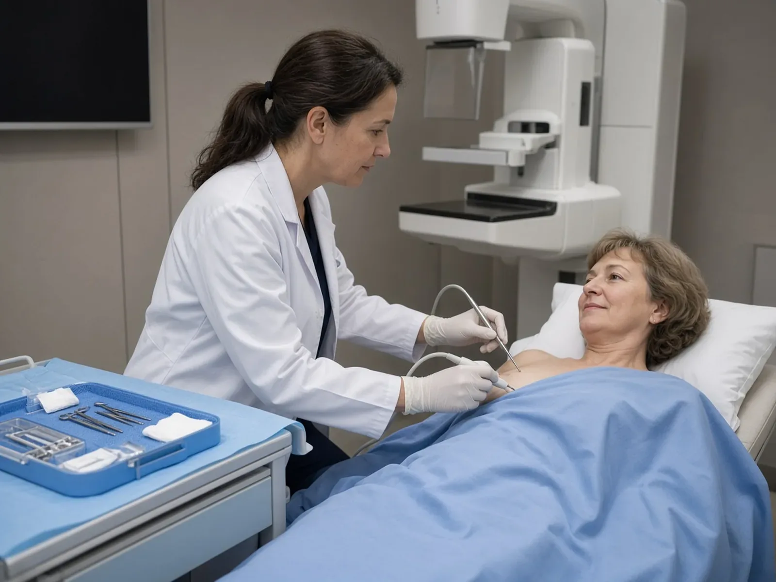
Localising a lesion that cannot be felt
The lesion generally cannot be felt. The radiologist therefore places a marker before the procedure: this is preoperative localisation. It guides the surgeon during the operation, to the lesion.
The hookwire is a thin metal wire, placed under local anaesthetic on the morning of the operation or the day before, which shows the surgeon where to operate. The magnetic clip can be placed several weeks before surgery.
The aim of surgery: to remove in one single block the localisation marker (hookwire or magnetic clip) and the surrounding lesion. An X-ray is performed at the end of the operation to check that everything has been removed.
The day of the procedure

On the day of the procedure, the surgeon removes the area that contains the lesion, localised beforehand if necessary, and keeps the rest of the breast. The specimen removed is then sent to the pathologist.
The incision and the scar
The incision and the scar depend on the size of the lesion and its location. In most cases, a single simple scar is enough — either directly over the lesion, or periareolar (around the areola) if the lesion is close to the nipple.
When the volume of tissue removed is significant relative to breast size, the remaining glandular tissue can be reshaped at the end of the procedure to restore a harmonious shape to the breast — this is called oncoplastic surgery. There is no standard incision — the technique is chosen on a case-by-case basis.
For a fibroadenoma, the incision is positioned as discreetly as possible — inframammary (in the fold below the breast) or axillary (in the armpit) whenever the topography allows. The line planned for you is shown at the consultation.
Practical arrangements
The practical arrangements (anaesthesia, duration, return home) are explained during your consultation. To get organised, see preparing for your surgery.
Margins: why they rarely matter here
The margins are the edges of the specimen: we look at whether abnormal cells touch them.
In cancer surgery, the question arises differently: see positive margins after lumpectomy.
Atypical lesions
An atypical ductal hyperplasia is never re-excised, even in case of involved margins or residual atypia on the specimen.
For radial scar, most often, the analysis confirms a radial scar, with or without atypia. In that case, there is usually no reason to operate again, even if the lesion touches the margins.
Lobular carcinoma in situ
Classic form: if the margins are involved, there is no re-excision. Pleomorphic or florid form: if the margins are involved, a further excision is recommended.
Phyllodes tumour
Phyllodes tumour is classified into three grades: benign, borderline, malignant. The principle is simple: remove the entire tumour en bloc, with a cuff of healthy tissue all around. The minimum margin recommended is ≥ 1 mm for benign phyllodes and ≥ 10 mm for borderline and malignant phyllodes.
For a benign phyllodes tumour with an involved margin, there is no further operation: surveillance by clinical examination and ultrasound is organised for 3 years. For a borderline or malignant phyllodes tumour, if the histological analysis shows insufficient margins for the given grade, a re-excision is proposed.
After the operation: result and recovery

After the operation, two things matter: the immediate recovery, and the result of the analysis of the specimen removed.
Recovery
Care, the return to your activities and the signs that should prompt you to call are described on the postoperative recovery page. The instructions specific to your procedure are given to you at the consultation.
The result of the final analysis
The whole area removed is analysed by the pathologist. Dr Zeitoun explains this result to you at a consultation after the operation.
Analysis of the specimen can also reveal a cancer that the biopsy had not seen: treatment is then that of a breast cancer; the choice of operation is explained in lumpectomy or mastectomy: how to choose.
Imaging surveillance
After the procedure, surveillance depends on the lesion removed. It relies on clinical examination and imaging, at set intervals.
Atypical ductal hyperplasia
A first mammo-ultrasound control at 6 months post-operatively (± magnetic resonance imaging (MRI) if the initial diagnosis was made or completed by MRI), then annual surveillance for at least 10 years by bilateral mammography + ultrasound (± MRI depending on context).
Lobular carcinoma in situ
A clinical breast examination every year; a two-view mammogram every year for 10 years, then every 2 years.
Radial scar
Surveillance by ultrasound and/or by mammogram is then planned for ten years, at intervals agreed with Dr Zeitoun.
Phyllodes tumour
- Benign. If a margin is involved: clinical examination and ultrasound for 3 years.
- Borderline. Clinical examination every 6 months for 2 years, then yearly up to 5 years. Annual mammography and ultrasound for 5 years.
- Malignant. Same schedule, with surveillance chest imaging.
Fibroadenoma
Fibroadenoma removed: systematic ultrasound check-up at 6 months, then return to standard screening rhythm according to age and risk factors. Fibroadenoma not removed: first check-up at 4 months then annual.

Would you like a surgical opinion before deciding whether or not to have your lesion removed?
Dr Zeitoun goes over the biopsy result and the imaging with you, then tells you what is appropriate in your case, operation or follow-up.
What you should expect from the consultation
The consultation is for reviewing your file, explaining the proposed option and its alternatives, and answering your questions. If a procedure is decided on, you are given a quote.
Documents to bring
- The written biopsy result, which gives the exact name of the lesion.
- Your mammograms, your ultrasounds and, if you have had one, your MRI, with their reports.
- Your personal and family history, and the list of your treatments.
For everything else, see preparing for your first consultation.
Where to consult
Consultations take place at the practice at 241 rue du Faubourg Saint-Honoré (Paris 8th) and at the Clinique Hartmann (Neuilly-sur-Seine). Surgery takes place at the Clinique Hartmann.
To find out about the training of the surgeon who sees you, see Dr Jérémie Zeitoun’s background.
Frequently asked questions
My biopsy says “benign”: why am I being offered an operation?
Because a benign lesion may be at risk: what we sampled is benign, but that sample is not enough to answer for the whole area. The operation checks that there is no cancerous lesion next to it, or responds to a radio-histological discordance. A fibroadenoma, entirely benign, may also be removed in four situations.
Is a lumpectomy for a benign lesion the same operation as for a cancer?
The principle is the same: removing an area of the breast and keeping the rest. The rules differ: no sentinel lymph node; no radiotherapy, except for pleomorphic lobular carcinoma in situ or lobular carcinoma in situ with necrosis and for borderline or malignant phyllodes tumour; and for an at-risk lesion such as atypical ductal hyperplasia, margins that are not clear do not lead to a second operation.
Can the lesion be monitored instead of removed?
Yes for a benign lesion: a fibroadenoma is monitored by default, a hamartoma is simply monitored. For an at-risk lesion, before any surveillance, a surgical or radiological excision is often required, discussed case by case at the multidisciplinary team meeting.
Can a vacuum-assisted biopsy replace surgery?
For B3 lesions, either a vacuum-assisted excision, performed by the radiologist, or surgery is proposed. For atypical ductal hyperplasia and phyllodes tumour, surgery remains preferred. These options are discussed at the multidisciplinary team meeting.
What happens if the analysis finally finds a cancer?
Across all B3 lesions, 10 to 30% of cancers are found on the surgical specimen. Management then becomes that of a breast cancer. Dr Zeitoun explains it to you at the consultation, with the report in hand.
If the margins are not clear, do I need further surgery?
For atypical ductal hyperplasia, classic lobular carcinoma in situ, a radial scar or a benign phyllodes tumour, no; for a benign phyllodes tumour, surveillance by clinical examination and ultrasound is organised for 3 years. For pleomorphic or florid lobular carcinoma in situ, a further excision is recommended. For a borderline or malignant phyllodes tumour, the minimum margin is ≥ 10 mm; if it is insufficient for the given grade, a re-excision is proposed.
What scar will there be after the operation?
It depends on the size of the lesion and its location. In most cases, a single simple scar is enough, periareolar (around the areola) if the lesion is close to the nipple; when the volume of tissue removed is significant relative to breast size, the breast is reshaped by oncoplastic surgery. For a fibroadenoma, the incision is inframammary or axillary whenever the topography allows.
What follow-up is there after the operation?
It depends on the lesion. Atypical ductal hyperplasia: first control at 6 months, then annual surveillance for at least 10 years. Benign phyllodes tumour with an involved margin: clinical examination and ultrasound for 3 years; borderline or malignant phyllodes tumour: over 5 years; fibroadenoma removed: ultrasound check-up at 6 months; fibroadenoma not operated on: first check-up at 4 months then annual.
What are Dr Zeitoun’s fees?
Dr Zeitoun practises in Sector 2 (non-OPTAM) and charges fees above the French Social Security rate for all consultations and procedures. Most international patients are not covered by French Social Security. A detailed quote is given during the consultation, before any procedure. If you are covered, the Assurance Maladie reimburses on the basis of the Social Security rate, and your health insurance may cover all or part of the additional fees, depending on your policy.
Sources
- Cancers et pathologies du sein : attitudes diagnostiques et thérapeutiques, protocoles de traitement, SENORIF 2025-2026. Management after a biopsy, lesions of uncertain malignant potential (B3), phyllodes tumour, margins, surveillance.
- Rubio I.T. et al., European guidelines for the diagnosis, treatment and follow-up of breast lesions with uncertain malignant potential (B3 lesions). European Journal of Surgical Oncology, 2024, 50, 107292. European recommendations cited by SENORIF: surgery or vacuum-assisted biopsy.
- Elfgen C. et al., Third International Consensus Conference on lesions of uncertain malignant potential in the breast (B3 lesions). Virchows Archiv, 2023, 483, 5-20. Consensus cited by SENORIF: vacuum-assisted excision or imaging follow-up in place of surgery.
- Lavoué V. et al., Clinical practice guidelines from the French College of Gynecologists and Obstetricians (CNGOF): benign breast tumors – short text. European Journal of Obstetrics & Gynecology and Reproductive Biology, 2016, 200, 16-23. French recommendations on benign breast tumours.
- Bishr M.K. et al., Contemporary management of phyllodes tumours of the breast: recommendations from the UK Association of Breast Surgery. British Journal of Surgery, 2025, 112(8), znaf152. Reference cited by SENORIF for the margins of phyllodes tumours.