


This article draws in particular on the European guidelines of ESGO, ESHRE and ESGE, the European societies of gynaecological oncology, reproductive medicine and gynaecological endoscopy.
A thick endometrium on ultrasound, or hyperplasia on your biopsy?
Bring the ultrasound, the MRI if one was done and the reports. Dr Jérémie Zeitoun, gynaecological cancer surgeon, sees you at the practice at 241 rue du Faubourg Saint-Honoré (Paris 8th) and at Clinique Hartmann (Neuilly-sur-Seine), where he operates.
What “endometrial hyperplasia” means
The endometrium is the lining that covers the inside of the uterus. The term hyperplasia is used when its glands multiply in a disorderly way and the lining thickens.

Hypertrophy on ultrasound, hyperplasia under the microscope
Ultrasound sees a thickness: this is called hypertrophy, or a thickened endometrium. It does not say what this lining contains.
Only analysis under the microscope by the pathologist, on a sample, can tell whether it is hyperplasia, a polyp or endometrial cancer. See understanding a pathology report.
Without atypia or with atypia: the distinction that changes everything
The World Health Organization classification distinguishes two forms:
- hyperplasia without atypia: the cells are numerous but look normal; it is a benign lesion;
- atypical hyperplasia, also called endometrioid intraepithelial neoplasia: the cells look abnormal; it is a precancerous lesion.
Why the lining thickens
The endometrium grows under the effect of oestrogens, the female hormones, when they are not balanced by progesterone. The most common situations are excess weight, cycles without ovulation (for example polycystic ovary syndrome), menopausal hormone therapy with oestrogens alone, and tamoxifen, a breast cancer treatment.
The tests before deciding
Most often, the story begins with heavy periods, bleeding between periods, or bleeding after the menopause. Sometimes the thickening is discovered by chance.

Pelvic ultrasound
Ultrasound, often done through the vagina, measures the thickness of the endometrium and looks for a polyp, a fibroid or an abnormality of the muscle of the uterus.

Pelvic MRI
Quite often, a pelvic MRI is done before the hysteroscopy, especially if endometrial cancer is suspected. It clarifies the thickening and looks at the muscle of the uterus. In women who have not reached the menopause, it can show adenomyosis.
Biopsy and hysteroscopy
An endometrial biopsy can be done during the consultation, with a thin cannula. It can, however, miss a lesion.
Hysteroscopy, which passes a small camera through the cervix into the uterus, makes it possible to see the whole cavity and to sample or remove the abnormal area precisely. For atypical hyperplasia, the American guidelines consider it the most reliable method for not missing an associated cancer.
Thick endometrium after the menopause: the most common question in consultation

This is the situation most often encountered: a postmenopausal woman in whom ultrasound shows a thickened endometrium. What should be done?
With bleeding
Any bleeding after the menopause must be investigated, even a simple trace. With a thickened endometrium, a hysteroscopy with sampling is offered. The full work-up is detailed in the article bleeding after the menopause.
Without bleeding, discovered by chance (asymptomatic patient)
The thickening is sometimes discovered during an ultrasound done for something else, without any symptom. The patient is asymptomatic.
Dr Zeitoun’s approach is then proactive: he always offers a hysteroscopy with sampling, to check under the microscope that there is nothing in particular. Guidelines vary from one country to another on this situation; this check makes it possible to remove the doubt with certainty.
What the result shows
Most often, the analysis shows a benign lining, a polyp or hyperplasia without atypia: the procedure has often been enough. If it shows atypical hyperplasia or a cancer, a hysterectomy is offered.
Hyperplasia without atypia: the uterus is preserved
The risk is low. According to the British college of obstetricians and gynaecologists, it is below 5% over 20 years, and most of these hyperplasias regress.
Correcting what can be corrected
The cause is treated first when possible: weight loss, adjustment of menopausal hormone therapy.
Before the menopause: treatment with progestogens
Before the menopause, progesterone and its derivatives, the progestogens, lead to a decrease in endometrial thickness. After the menopause, they are not offered. They can be given:
- by a levonorgestrel IUS (hormonal coil), which releases the progestogen directly into the uterus; it makes hyperplasia regress better than tablets, with fewer side effects;
- by tablets, continuously, for at least 6 months.
A check of the endometrium then verifies that the hyperplasia has regressed.
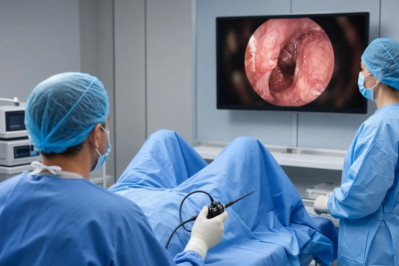
Operative hysteroscopy with endometrial resection
Operative hysteroscopy makes it possible to remove the thickened lining: this is endometrial resection, a resection of the superficial layer of the endometrium. It treats and gives, at the same time, all of the tissue to be analysed.
How to choose
Before the menopause, there is no treatment to be preferred from the outset. The choice between progestogens and operative hysteroscopy always depends on your age and your situation: wish for pregnancy, bleeding, other treatments. It is decided with you in consultation.
After the menopause, progestogens are not offered: operative hysteroscopy with endometrial resection is the option chosen.
When hysterectomy is discussed
For hyperplasia without atypia, hysterectomy is not the first-line treatment. It is discussed if atypia appears, if the hyperplasia does not regress after 12 months of treatment, if it recurs, if the bleeding persists, or if you request it after being informed.
You have hyperplasia without atypia and you want to know whether a hysteroscopy is enough?
Dr Zeitoun goes over the ultrasound, the biopsy and your symptoms with you, and explains the options that preserve the uterus.
On tamoxifen: a known and monitored thickening

Tamoxifen, prescribed after breast cancer, stimulates the endometrium. It often leads to a so-called glandulocystic hypertrophy, that is, with small fluid-filled cavities, well known to radiologists.
Monitoring
This appearance is monitored by ultrasound. Report any bleeding to your doctor.
When the endometrium keeps thickening
If the endometrium thickens progressively, an operative hysteroscopy with endometrial resection is offered. There is no reason to perform a hysterectomy, all the more so if the analysis confirms a lesion without atypia. Tamoxifen can be continued, in liaison with your oncologist.
Atypical hyperplasia: hysterectomy is the standard treatment
Two reasons explain this approach. First, a review of eleven studies finds a cancer already present in 32.6% of cases on average. Second, the risk of progression to cancer is about 8% per year.
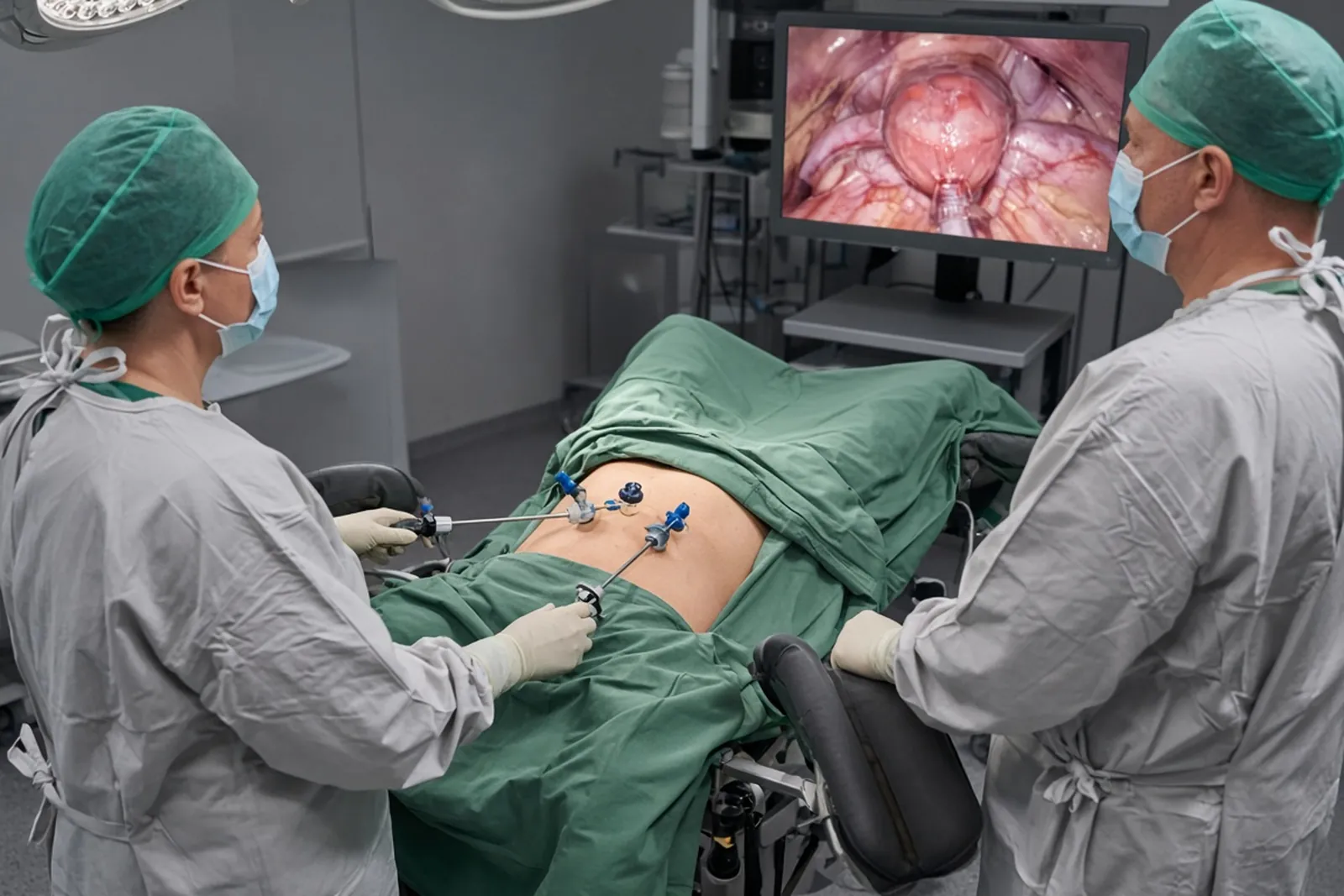
What the operation involves
Hysterectomy removes the uterus and the cervix. The guidelines advise against leaving the cervix (so-called subtotal hysterectomy) in this situation.
The operation is most often performed by laparoscopy, through small incisions, with a short hospital stay. Dr Zeitoun operates at Clinique Hartmann.
The ovaries and the tubes
The tubes are removed at the same time. After the menopause, the ovaries are removed or preserved depending on the case; the decision is made with you before the operation. See surgery of the ovaries and tubes.
Analysis of the uterus
The removed uterus is analysed in full. If it contains a cancer, the next steps are discussed at a multidisciplinary team meeting, which brings together the surgeon, radiologist, pathologist, oncologist and radiation oncologist. See stage 1 endometrial cancer and pelvic sentinel node.
Atypical hyperplasia and wish for pregnancy

A woman who wishes to become pregnant can, in some cases, keep her uterus despite atypical hyperplasia. Guidelines on this point are rare and imprecise, because no randomised controlled trial has been carried out. What follows takes up the British reference guideline and the protocol of a French national reference centre.
Before deciding
You are first informed of the risk that a cancer is already present, and of the risk of progression to cancer.
The work-up seeks to rule out an invasive endometrial cancer or an associated ovarian cancer. The French reference centre requires at least a pelvic MRI, as well as a hysteroscopy and a laparoscopy under general anaesthetic to guide the samples.
A fertility assessment is carried out, and you are referred to a fertility specialist. The results are reviewed at a multidisciplinary team meeting, which sets the treatment and the monitoring.
This choice requires accepting prolonged and rigorous monitoring.
Treatment
Treatment is based on progestogens. The British guideline proposes the levonorgestrel IUS (hormonal coil) as first-line treatment, and progestogen tablets as an alternative. The French reference centre points out that there is no consensus on the choice of progestogen, its dose, its route of administration and its duration.
The same centre specifies that no extensive resection of the endometrium should be performed as part of this treatment, because it can compromise fertility and pregnancies.
Monitoring
Endometrial biopsies are done every 3 months, until two consecutive negative biopsies are obtained. In the protocol of the French reference centre, the first check by hysteroscopy with sampling takes place after 3 months of treatment.
Pregnancy
Pregnancy is attempted only after the atypia has disappeared on at least one sample. The progestogen is then stopped, and pregnancy is sought as soon as possible. Medically assisted reproduction may be offered.
If the atypia persists or progresses
In the event of progression, a hysterectomy is indicated. In the event of persistence, you are offered, after discussion, either a hysterectomy or a second medical treatment.
When pregnancy is no longer wished
A hysterectomy is then offered, because of the high risk of recurrence.
When the thickening is explained by adenomyosis
In some women who have not reached the menopause, hypertrophy of the endometrium is part of adenomyosis: the lining infiltrates the muscle of the uterus. The diagnosis is made after the hysteroscopy and a pelvic MRI.
Close to the menopause
Dr Zeitoun often offers an operative hysteroscopy with superficial endometrial resection. Treatment with progestogens is also possible. The advantage of hysteroscopy is to obtain a reliable histological sample, that is, tissue analysed under the microscope.
In a young woman who wishes to become pregnant
A trial treatment with progestogens is offered, as microprogestogens (low doses) or macroprogestogens (higher doses). A reassessment at 2 or 3 months checks the decrease in endometrial thickness. Otherwise, an operative hysteroscopy with endometrial resection is offered.
Some macroprogestogens, such as chlormadinone acetate or nomegestrol acetate, increase the risk of meningioma, a most often benign tumour of the membranes covering the brain. The French National Agency for Medicines Safety (ANSM) asks that they be prescribed at the minimum effective dose, for the shortest possible time, with monitoring by brain MRI when treatment continues beyond one year.
You are hesitating between conservative treatment and hysterectomy, or you are planning a pregnancy?
Dr Zeitoun explains what each option brings you, what follow-up it requires, and what he recommends in your case.
Hysteroscopy or hysterectomy: the decision table
| Situation | What is most often offered |
|---|---|
| Thick endometrium after the menopause, with bleeding | Hysteroscopy with sampling |
| Thick endometrium after the menopause, without bleeding (asymptomatic) | Hysteroscopy with sampling, always offered by Dr Zeitoun |
| Hyperplasia without atypia, before the menopause | Progestogens (levonorgestrel IUS (hormonal coil) or tablets) or operative hysteroscopy with endometrial resection, depending on age and situation; uterus preserved |
| Hyperplasia without atypia, after the menopause | Operative hysteroscopy with endometrial resection; no progestogens |
| Thickening on tamoxifen | Monitoring by ultrasound; endometrial resection if it thickens; no hysterectomy if the analysis shows no atypia |
| Adenomyosis close to the menopause | Operative hysteroscopy with superficial endometrial resection most often (reliable histological sample), or progestogens |
| Adenomyosis and wish for pregnancy | Trial of progestogens, check at 2-3 months, endometrial resection if endometrial thickness does not decrease |
| Atypical hyperplasia, no wish for pregnancy | Total hysterectomy, most often by laparoscopy |
| Atypical hyperplasia and wish for pregnancy | Progestogens (levonorgestrel IUS (hormonal coil) as first-line treatment according to the British guideline), biopsies every 3 months, pregnancy after the atypia has disappeared; hysterectomy when pregnancy is no longer wished |
The procedure in practice
Operative hysteroscopy
It is performed through the natural passages, without a scar, most often as a day case, under general or regional anaesthetic depending on the case. Recovery is generally simple: mild pain comparable to period pain and light bleeding for a few days. See hysteroscopy.
Hysterectomy
It is most often performed by laparoscopy, with a short hospital stay. See hysterectomy, preparing for your operation and postoperative recovery.
What you should expect from the consultation
The consultation is used to go over the ultrasound, the MRI, the biopsy and your treatments, particularly hormonal ones. Dr Zeitoun examines you, explains the exact meaning of the result and compares the options with you.
Bring the images, the reports and the list of your treatments. See preparing for your first consultation, benign surgery of the uterus and Dr Zeitoun’s background.
Frequently asked questions
My endometrium is thick after the menopause, without bleeding: what should be done?
Even without any symptom, Dr Zeitoun always offers a hysteroscopy with sampling, to check under the microscope that there is nothing in particular. If there is bleeding, this investigation is also necessary. See bleeding after the menopause.
Is endometrial hyperplasia a cancer?
No. Without atypia, it is a benign lesion, with a low risk of progression. With atypia, it is a precancerous lesion, and a cancer is already present in about one third of cases.
Does the uterus have to be removed for hyperplasia without atypia?
In general, no. Before the menopause, treatment with progestogens or an operative hysteroscopy with endometrial resection, chosen according to your age and your situation, make it possible to preserve the uterus. After the menopause, operative hysteroscopy is what is offered. Hysterectomy is discussed in the event of failure, recurrence or persistent bleeding.
Why hysterectomy for atypical hyperplasia?
Because a cancer is often already present, and the risk of progression is high. Hysterectomy treats the lesion and makes it possible to analyse the whole uterus.
Can you become pregnant after atypical hyperplasia?
Sometimes, yes. Treatment with progestogens can be offered, with check biopsies every 3 months. Pregnancy is attempted after the atypia has disappeared, and a hysterectomy is offered when pregnancy is no longer wished.
Can the hormonal coil treat hyperplasia?
Yes, before the menopause. The levonorgestrel IUS (hormonal coil) is an effective treatment for hyperplasia without atypia. The British guideline also proposes it as first-line treatment in the conservative treatment of atypical hyperplasia. After the menopause, it is not offered.
I am taking tamoxifen and my endometrium is thick: is it serious?
Most often, no. Tamoxifen thickens the endometrium in a known way. It is monitored by ultrasound; if it keeps thickening, an operative hysteroscopy with endometrial resection is offered, without hysterectomy if the analysis shows no atypia. See hormone therapy.
What is the link with adenomyosis?
Before the menopause, a thick endometrium can be part of adenomyosis. Pelvic MRI helps to show it, and treatment depends on your age and your wish for pregnancy.
Is operative hysteroscopy painful?
It is performed under general or regional anaesthetic depending on the case, most often as a day case. Recovery is generally simple, with pain comparable to period pain for a few days.
What are Dr Zeitoun’s fees?
Dr Zeitoun practises in Sector 2 (non-OPTAM) and charges fees above the French Social Security rate for all consultations and procedures. Most international patients are not covered by French Social Security. A detailed quote is given during the consultation, before any procedure. If you are covered, the Assurance Maladie reimburses on the basis of the Social Security rate, and your health insurance may cover all or part of the additional fees, depending on your policy.
Sources
- Rodolakis A. et al. — ESGO/ESHRE/ESGE Guidelines for the fertility-sparing treatment of patients with endometrial carcinoma. Human Reproduction Open, 2023, no. 1, hoac057 (doi: 10.1093/hropen/hoac057; PMID: 36756380). European guidelines devoted to endometrial cancer; they state that no data distinguish the management of cancer from that of atypical hyperplasia.
- Royal College of Obstetricians and Gynaecologists / British Society for Gynaecological Endoscopy — Management of Endometrial Hyperplasia, Green-top Guideline No. 67, 2016. Hyperplasia without atypia: risk of progression to cancer below 5% over 20 years; levonorgestrel IUS more effective than tablets; treatment for at least 6 months; hysterectomy not offered as first-line treatment. Atypical hyperplasia and wish for pregnancy: information on the risk of cancer, work-up, multidisciplinary meeting, levonorgestrel IUS as first-line treatment, biopsies every 3 months until two negative biopsies, pregnancy after regression, hysterectomy when pregnancy is no longer wished.
- Hôpital Bichat – Claude-Bernard, centre de référence PREFERE — Hyperplasie atypique : principes du traitement conservateur (in French) (page updated on 17/07/2026). Work-up (pelvic MRI, hysteroscopy and laparoscopy, fertility assessment), progestogens with no consensus on the molecule and the duration, check at 3 months, management according to the response, no extensive endometrial resection, hysterectomy as the last step.
- American College of Obstetricians and Gynecologists — Management of Endometrial Intraepithelial Neoplasia or Atypical Endometrial Hyperplasia, Clinical Consensus No. 5. Obstetrics & Gynecology, 2023, 142(3) (PMID: 37590985). Hysterectomy as the standard treatment; hysteroscopy with sampling as the most reliable method to look for an associated cancer; no subtotal hysterectomy; associated cancer in 32.6% of cases (review of 11 studies) and progression of about 8% per year.
- Doherty M.T. et al. — Concurrent and future risk of endometrial cancer in women with endometrial hyperplasia: a systematic review and meta-analysis. PLoS One, 2020, 15(4), e0232231 (doi: 10.1371/journal.pone.0232231; PMID: 32343732). Eleven studies: associated cancer in 32.6% of atypical hyperplasias; progression to cancer of about 8% per year.
- Agence nationale de sécurité du médicament (ANSM) — Progestatifs et risque de méningiome : recommandations pour limiter ce risque (in French). Minimum effective dose, shortest duration, annual reassessment, brain MRI depending on the duration of treatment.
- Catena U. et al. — A practical guideline on the fertility-sparing treatment of patients with endometrial carcinoma and atypical endometrial hyperplasia. International Journal of Gynecology & Obstetrics, 2025 (doi: 10.1002/ijgo.70059). Reproductive medicine opinion before conservative treatment; hysterectomy recommended once the pregnancy plan has been completed.
Article written by Dr Jérémie Zeitoun, breast and gynaecological cancer surgeon.