Bleeding after the menopause, a diagnosis of stage 1 uterine cancer: news that is hard to hear, yet one that carries an excellent prognosis when managed properly. Why is surgery the standard treatment? What does the operation involve? What do the sentinel lymph node and the new FIGO 2023 classification add? This article takes stock.
Dr Zeitoun sees patients in consultation to review your file, explain the indication for surgery, the technique and the care pathway, and to support you from diagnosis through to recovery. A second opinion is available.
Endometrial cancer — also called cancer of the body of the uterus — arises in the lining of the inside of the uterus. It is the most common gynaecological cancer in France (ahead of ovarian cancer), with nearly 8,000 new cases every year. It occurs most often after the menopause and, fortunately, is usually diagnosed at an early stage because it shows itself early through a recognisable warning sign.
The term stage 1 refers to a tumour confined to the body of the uterus, with no spread to the cervix, the lymph nodes or distant organs. Though hard to hear, this is a diagnosis that carries an excellent prognosis when managed properly.
This article explains why surgery is the standard treatment at stage 1, what the operation involves, what the sentinel lymph node and minimally invasive techniques add, how recovery unfolds, and how the new FIGO 2023 classification and molecular profiles now personalise care. For the disease as a whole, see also our dedicated page on endometrial cancer.
Indication, minimally invasive technique, sentinel lymph node, pre- and post-operative pathway: a direct consultation with the surgeon.
At stage 1, the tumour remains confined to the body of the uterus. Understanding how the diagnosis is made, and what the analysis reveals, makes it easier to approach the question of treatment with greater peace of mind.
The most common symptom — and often the sign that leads to the diagnosis — is abnormal vaginal bleeding: any bleeding after the menopause, or abnormally heavy or prolonged periods in a woman who is not menopausal. Any postmenopausal bleeding should prompt a consultation without delay. To find out more, read our dedicated article on bleeding after the menopause.
Although endometrial cancer occurs most often after the menopause, gynaecological cancers do exist — exceptionally — in young women. This is why abnormal bleeding should never be dismissed, even before the menopause: in the great majority of cases the cause is benign (see benign conditions of the uterus), but only a medical opinion can confirm this.
The approach combines several complementary tests, from the simplest to the most precise.
Transvaginal ultrasound is the first-line examination. It measures the thickness of the endometrium: a thin endometrium in a postmenopausal woman makes cancer very unlikely.
An office biopsy (Cornier pipelle) is a simple first step, but it is often of little help, or even normal: on its own it can only rarely confirm the diagnosis. If doubt persists, a hysteroscopy with directed biopsies (or even a curettage) is needed for a reliable diagnosis.
The reference examination for local staging. It assesses the depth of invasion of the uterine muscle (myometrium) and the state of the lymph nodes.
👉 Pelvic MRI remains the reference examination for local staging. For distant staging, a PET-CT scan may be performed as a matter of course to look for distant spread, particularly in higher-risk cases; CA 125 is the reference biological marker used in this setting. These tests are ordered on a case-by-case basis and interpreted by the team (2026 data).
Historically, stage 1 was divided according to the depth of myometrial invasion: stage 1A (invasion of less than half the thickness of the muscle) and stage 1B (invasion of half or more). This distinction remains useful.
But since 2023, the international FIGO classification has changed profoundly: it is no longer based on the anatomy of the tumour alone. It now incorporates the cell type (histology), invasion of the small vessels (LVSI) and, above all, the molecular biology of the tumour. This is a major advance — more refined but also more complex and debated — that opens the way to more personalised medicine.
Analysis of the tumour allows it to be classified into one of four groups, each with a different prognosis and different treatment implications.
Excellent prognosis. These tumours can sometimes justify de-escalating additional treatments, even at an early stage.
A defect in the DNA repair system. Intermediate prognosis; an important profile because it is sensitive to immunotherapy in advanced disease.
Intermediate prognosis. This is the most common group: the tumour shows none of the three abnormalities that characterise the other profiles.
A more guarded prognosis, which may lead to intensifying treatment even at an early stage.
In practice, your molecular group influences two things: the prognosis and the intensity of the treatment offered after surgery. Here, in plain language, is what each profile implies.
| Profile | In plain terms | Prognosis | Treatment intensity |
|---|---|---|---|
| POLE mutated | A particular "signature" in the tumour's DNA, fairly rare. | Excellent — the most favourable of the four. | Often de-escalated: at an early stage, radiotherapy or chemotherapy can sometimes be avoided. |
| MMR deficient (dMMR) | The tumour repairs its DNA poorly. Sometimes linked to Lynch syndrome (hereditary risk), which should be checked for. | Intermediate. | Tailored to risk. A key profile because it is sensitive to immunotherapy if the disease becomes advanced or recurs. |
| Non-specific (NSMP) | The most common group: none of the three other abnormalities. | Intermediate, most often favourable at stage 1. | Decided according to grade, muscle invasion and LVSI: surveillance or brachytherapy in most cases. |
| p53 abnormal (p53abn) | An abnormality of a protein that controls cells; more aggressive tumours. | More guarded. | Often intensified, even at an early stage (radiotherapy and/or chemotherapy). |
👉 In practice, two tumours of identical size may now be managed differently according to their molecular profile. This recent approach, more refined but also more complex, requires access to specialised testing; your file is always discussed by the team to take this into account. For the overall picture of the disease, see the dedicated page on endometrial cancer.
Any postmenopausal bleeding warrants a consultation without delay. If endometrial cancer has been diagnosed or suspected, Dr Zeitoun will see you to discuss the work-up, the indication for surgery and the care pathway.
For stage 1 endometrial cancer, the question of surgery rarely arises: surgery is the standard treatment, recommended by French and international learned societies (INCa, CNGOF, ESGO-ESTRO-ESP). It plays both a curative role — it removes the tumour — and an essential diagnostic role in guiding any additional treatment.
Removing the uterus eliminates the source of the cancer. It is the most effective way to achieve a definitive cure at stage 1.
Analysis of the surgical specimen confirms the stage, grade, vascular invasion and molecular profile of the tumour.
These results determine whether or not to add brachytherapy, radiotherapy or chemotherapy.
Standard surgery comprises three complementary steps.
In some young women who wish to become pregnant, with a stage 1A, low-grade (grade 1) tumour and no myometrial invasion, temporary conservative treatment with hormones (progestogens or a levonorgestrel intrauterine device) may be discussed at a multidisciplinary team meeting (MDT).
The conditions are specific: stage IA, grade 1, an endometrioid tumour with no myometrial invasion and no sign of spread on MRI, a decision approved at the MDT, and acceptance of close surveillance by hysteroscopy (follow-up biopsies, typically every 3 to 6 months). Regular hysteroscopy is essential to check the response.
This treatment is not the rule: it is an exceptional option, carrying a higher risk of recurrence. Surgery remains recommended once the pregnancy plan has been fulfilled, or if hormone treatment does not work. An important nuance: in the case of a dMMR tumour or Lynch syndrome, fertility preservation is more delicate and must be discussed with particular caution. If this situation applies to you, talk about it openly at your consultation.
Before surgery, it is natural to want some points of reference. Here are useful questions to note down and take to your consultation. For the practical organisation, see also our guide to preparing for your operation.
Surgical route, sentinel lymph node, length of hospital stay, recovery: Dr Zeitoun answers all your questions in consultation.
Surgery for endometrial cancer has changed considerably over the past twenty years. Today, minimally invasive techniques are preferred: they offer oncological outcomes equivalent to open surgery, with major benefits for recovery. This equivalence has been demonstrated by large comparative trials (GOG LAP2, LACE trial), which confirmed lower morbidity and better quality of life without any compromise on disease control.
The preferred approach for the great majority of patients. Under general anaesthesia, 3 to 5 small incisions of less than a centimetre, a high-definition camera and fine instruments allow the surgery to be performed with precision. Hospital stay often 1 to 3 days, reduced pain, return to light activity in 2 to 4 weeks.
Robotic surgery (da Vinci-type robot) exists and is expanding. The literature — meta-analyses and a randomised trial — shows oncological outcomes equivalent to those of laparoscopy: the robot mainly brings technical advantages (fewer conversions, useful in cases of obesity), with no oncological superiority. Dr Zeitoun, for his part, operates by laparoscopy.
Surgery through an abdominal incision remains indicated in certain cases: a very large uterus, extensive adhesions, contraindications to the minimally invasive route or certain advanced forms. The decision is made on a case-by-case basis.
👉 Dr Zeitoun performs minimally invasive surgery by laparoscopy, an expertise developed in reference centres such as Institut Gustave Roussy and Institut Curie. Before the operation, find out how to prepare for your surgery.
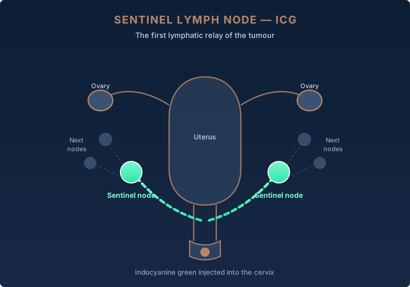
The sentinel lymph node technique has transformed surgery for endometrial cancer.
A fluorescent tracer — indocyanine green (ICG) — is injected into the cervix. Using an infrared camera, it makes it possible to identify the first node or nodes that drain the tumour. These nodes are removed and analysed.
When the sentinel nodes contain no cancer cells, an extended lymph node dissection is generally not necessary.
Everything takes place during the operation, under general anaesthesia: you feel nothing and have nothing in particular to prepare for this step.
The benefit for you: by removing only the truly useful nodes, reliable information is obtained while greatly reducing the risk of lymphoedema — that chronic, disabling swelling of the legs that could follow the extended dissections of the past.
Validated by prospective studies (the FIRES trial in particular), the sentinel lymph node technique has largely replaced extended lymph node dissection in early stages.
👉 The surgical decision is personalised. Your file is systematically presented at a multidisciplinary team meeting (MDT), bringing together surgeon, oncologist, radiation oncologist, radiologist and pathologist. It is this collective discussion that ensures the treatment best suited to your situation.
Understanding what happens after the operation makes it possible to prepare calmly. Recovery is generally well tolerated, especially with minimally invasive surgery, and most patients return to a normal life within a few weeks.
After the operation, you are monitored in the recovery room. Pain is managed with appropriate painkillers. You can usually drink that same evening.
Early mobilisation from the next day, gradual resumption of eating. Compression stockings and sometimes anticoagulants prevent thrombosis. With the minimally invasive route, discharge often takes place after 1 to 3 days.
Relative rest in the first days, gradual return to light activities. No heavy lifting for 4 to 6 weeks. Sexual activity resumes after the follow-up consultation.
The final pathology results, including the molecular profile, are available in 2 to 3 weeks. Your file is presented again at the MDT to decide on any additional treatments.
Every woman recovers at her own pace and these markers apply mainly to minimally invasive surgery; they give you an idea of the usual trajectory.
| When | What happens | What you can do |
|---|---|---|
| Day of surgery (D0) | Waking up in the recovery room, pain controlled by painkillers, a drip. Sometimes a small urinary catheter, removed quickly. | Drink, then eat lightly in the evening. Move your legs in bed. |
| Day 1 to 3 | Early mobilisation from the next day, resumption of eating, compression stockings ± anticoagulants against thrombosis. Discharge often at this stage. | Walk on the ward then at home, wash yourself, climb a few steps. |
| Weeks 1 to 2 | Normal tiredness, some slight vaginal discharge possible. Incisions of a few millimetres. Simple painkillers. | Gentle daily walking. No heavy lifting, no driving while in pain or on strong painkillers. |
| Weeks 3 to 6 | Energy returning, internal healing under way. Follow-up consultation during this period. | Return to light activity and often to work. Still no heavy lifting or prolonged baths. |
| 1 to 2 months | Final pathology results (including molecular profile) available; file presented again at the MDT. | Gradual return to sport and intimate life once the surgeon gives the go-ahead. |
If you were not yet menopausal, removing the ovaries induces an immediate menopause. The symptoms — hot flushes, vaginal dryness, sleep disturbance — can be pronounced because they appear abruptly. The question of hormone replacement therapy (HRT) is discussed on a case-by-case basis at the MDT: it is neither systematically allowed nor systematically forbidden. The decision takes into account the type and profile of the tumour, your symptoms and any contraindications. Conversely, oestrogen-only treatment (without a progestogen) is among the risk factors for this cancer, which explains the caution.
This is never a decision to be taken alone or in a hurry: other solutions (local treatments for vaginal dryness, non-hormonal measures) exist and will be discussed with you.
The need for treatment after the operation depends on the final results and on the risk profile for recurrence, which now incorporates molecular biology.
👉 The biology modulates these decisions: an early-stage POLE mutated tumour may justify de-escalating treatment, whereas a p53 abnormal tumour tends to lead to intensifying it.
Recent advances (the RUBY trial with dostarlimab and NRG-GY018 with pembrolizumab) have shown a major benefit of immunotherapy, particularly in MMR deficient tumours.
These treatments currently concern advanced or recurrent disease, and are not part of the standard management of stage 1.
👉 Good to know: most recurrences occur in the first years. The prognosis of a properly treated stage 1 endometrial cancer is excellent, and now also depends on the molecular group of the tumour.
The questions that come up most often in consultation. If yours is not here, do not hesitate to ask it at your appointment — or to Sophie, the site's assistant, at the bottom right.
Yes, stage 1 endometrial cancer is most often curable. Diagnosed at an early stage, it carries an excellent prognosis: 5-year survival most often exceeds 90% for localised disease. Surgery is the cornerstone of treatment. The prognosis is now refined by the molecular classification (POLE mutated tumours have an excellent prognosis, p53 abnormal tumours a more guarded prognosis).
The FIGO 2023 classification no longer relies solely on the anatomy of the tumour: it incorporates the histological type, vascular invasion (LVSI) and, above all, the molecular biology (POLE mutated, MMR deficient, p53 abnormal, or non-specific profile). Two tumours of the same size can therefore be classified differently according to their profile, which allows treatments to be tailored more precisely — sometimes by de-escalating them.
In the vast majority of cases, surgery is the standard treatment. Patients who cannot be operated on may receive radiotherapy, with less good results. Conservative treatment with progestogens is reserved for very selected cases of young women wishing to preserve their fertility, under strict surveillance and after a decision at the MDT.
Standard surgery combines removal of the whole uterus — body and cervix (total hysterectomy) — and removal of both ovaries and both tubes (bilateral salpingo-oophorectomy). This is most often supplemented by exploration of the lymph nodes using the sentinel lymph node technique. In the great majority of cases it is performed by a minimally invasive route (laparoscopy or robot).
The sentinel lymph node is the first lymphatic relay draining the tumour. It is identified by injecting a fluorescent tracer (indocyanine green), then removed for analysis. This technique has largely replaced extended dissection in early stages: it gives reliable information about the nodes while greatly reducing the risk of lymphoedema (chronic swelling of the legs).
After minimally invasive surgery (laparoscopy or robot), the hospital stay is short, often 1 to 3 days or even day-case depending on the centre. After open surgery (laparotomy) it is longer. Return to light activity usually takes 2 to 4 weeks after minimally invasive surgery.
This depends on analysis of the surgical specimen and the molecular profile. Many low-risk stage 1 patients need no additional treatment (surveillance alone). Others receive vaginal brachytherapy (intermediate risk) or external radiotherapy ± chemotherapy (high risk). Immunotherapy is reserved for advanced or recurrent disease, mainly MMR deficient; it is not part of the standard treatment of stage 1.
Like any operation, surgery carries risks: bleeding, infection, injury to neighbouring organs (ureters, bladder, bowel) and anaesthetic risk. In the long term: surgical menopause if you were not yet menopausal, possible vaginal dryness, and a small risk of lymphoedema (reduced by the sentinel lymph node). Your surgeon explains these points in detail at the pre-operative consultation.
This article is for information purposes and does not replace a medical consultation. Treatment decisions are made on a case-by-case basis at a multidisciplinary team meeting.
To go further in understanding endometrial cancer and its surgical management.
Everything you need to know about cancer of the body of the uterus: diagnosis, stages, treatments and management by Dr Zeitoun.
ProcedureSurgical routes (laparoscopy, vaginal, laparotomy), post-operative course, recovery — everything you need to know.
HubCervix, endometrium, ovary, vulva: an overview of the gynaecological cancers managed and the techniques offered.
Do not face this news alone. Dr Jérémie Zeitoun sees patients in consultation at his practice in the 8th arrondissement of Paris and at Clinique Hartmann in Neuilly-sur-Seine to answer all your questions, review your file and offer you the best personalised treatment strategy.