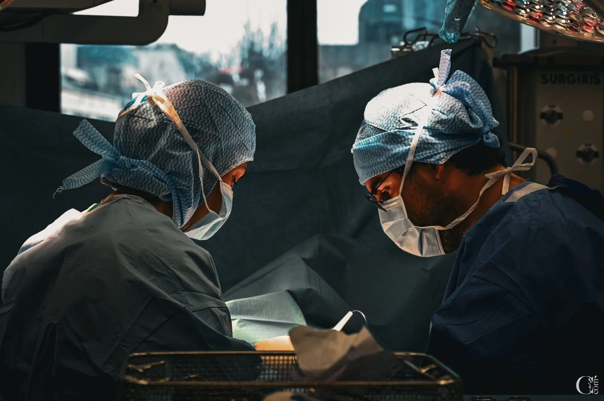
A benign outgrowth of the cervix, often responsible for bleeding. Removal by forceps or operative hysteroscopy, with systematic histology.
Author and medical review: Dr Jérémie Zeitoun, cancer surgeon (gynaecology & breast) — RPPS 10101463296 — trained at Institut Gustave Roussy, Institut Curie and Centre François Baclesse. See full background →
Last updated: · Sources: CNGOF, HAS, NHS, RCOG. References at the bottom of the page.
A cervical polyp is an outgrowth that develops on the surface of the cervix or inside the endocervical canal. It appears as a soft, bright-red, rounded or elongated lesion, most often pedunculated (attached to the cervix by a thin stalk) or, less often, broad-based. Cervical polyps are, in the overwhelming majority of cases, benign; a malignant nature remains possible, though very exceptional.
Cervical polyps range in size from a few millimetres to several centimetres. Most are under 1 cm and remain undetected for years. Larger polyps protrude from the cervical os and are visible at speculum examination.
Cervical polyps are a common finding, often discovered incidentally during a routine examination. They mainly affect women between 40 and 60, but can occur at any age, including after menopause. The overwhelming majority remain benign throughout life; a small risk of adenomatous or atypical polyp nevertheless justifies systematic removal with histopathological analysis.
The two are frequently confused. A cervical polyp arises from the endocervical canal or the exocervix and may be visible at speculum examination. An endometrial polyp develops inside the uterine cavity and is only detected by ultrasound or hysteroscopy. The two may coexist in the same patient — a key reason for offering a diagnostic hysteroscopy in theatre when a high or endocervical polyp is confirmed, so that the uterine cavity can be assessed in the same sitting.
The exact mechanisms behind cervical polyp formation are not fully understood. They probably result from a localised proliferation of the cervical mucosa under the influence of several hormonal and inflammatory factors.
Prolonged oestrogenic stimulation promotes cervical mucosal proliferation. It explains why cervical polyps are more frequent in perimenopausal women and in patients with obesity. Hormone replacement therapy can also contribute.
Chronic cervicitis, recurrent infection or mechanical irritation can lead to local hyperplasia and polyp formation. This is one of the reasons polyps bleed readily on contact.
Cervical polyps are more common in multiparous women and after age 40. Estimated prevalence in the general population is between 2% and 5%. They are rarer in young nulliparous women.
Diabetes, obesity, tamoxifen (hormonal therapy for breast cancer) and certain hormone replacement regimens are associated with an increased risk of cervical and endometrial polyps. Systematic screening is warranted in these situations.
Cervical polyps are not linked to HPV infection. Unlike cervical dysplasia (CIN), which results from persistent human papillomavirus infection, polyps are mechanical and inflammatory lesions. However, they can coexist with HPV-related lesions, which is why a colposcopic examination is often included in the consultation.
Source: CNGOF 2024
Many cervical polyps are entirely asymptomatic and found incidentally during a gynaecological examination or a cervical screening. When symptoms do occur, bleeding is the most characteristic feature.
Any bleeding outside periods — whether after intercourse, between cycles or after menopause — warrants a gynaecological consultation. Speculum examination often makes the diagnosis immediately when the polyp protrudes through the cervical os. What matters most is not to dismiss these bleeds, however minor: they can reveal a benign polyp, but may also, more rarely, point to a more concerning lesion that needs to be diagnosed early.
Source: CNGOF 2024
Cervical polyp diagnosis is clinical in most cases: speculum examination alone often shows a bright-red lesion emerging from the cervical canal. Further investigations confirm the diagnosis, map the lesion, and rule out associated pathology.
The first diagnostic step. The polyp appears as a bright-red outgrowth, pedunculated or sessile, emerging from the cervical os. Its size, colour, base of insertion and contact bleeding are noted.
Examination of the cervix under magnification after application of acetic acid and Lugol's iodine. It helps rule out an associated dysplastic lesion and better characterises the polyp.
Assesses for an associated endometrial polyp, fibroid or uterine abnormality. Often performed before hysteroscopy when an endocervical polyp is suspected or in any patient over 40.
The reference examination for the endocervical canal and uterine cavity. In Dr Zeitoun's practice, diagnostic hysteroscopy is performed in the operating theatre, combined if needed with polyp removal in the same sitting.
Over 40, the workup is systematically more thorough. The risk of an associated endometrial polyp, benign uterine pathology (fibroid, hyperplasia) or — more rarely — a malignant lesion justifies going beyond speculum examination. Dr Zeitoun typically requests a pelvic ultrasound and very often a hysteroscopy, diagnostic or operative depending on the case.
Source: HAS · CNGOF 2024
The removal technique depends on three criteria: the size of the polyp, the width of its base and the age of the patient. Dr Zeitoun systematically tailors the strategy to each situation.
The choice is not arbitrary. A thin, pedunculated, small and clearly visible polyp in a younger patient is ideal for forceps removal in the clinic — a few seconds, no anaesthesia, no time off. Conversely, a polyp that is broad-based, large, high in the canal or discovered in a patient over 40 warrants removal in theatre by operative hysteroscopy. This approach ensures complete resection of the stalk and detects an associated endometrial polyp, which is common in this age group.
In Dr Zeitoun's practice, diagnostic hysteroscopy is performed in the operating theatre rather than in the outpatient clinic. This organisation allows optimal patient comfort, complete assessment of the canal and cavity, and removal in the same sitting if a lesion is identified. Some colleagues with appropriate equipment perform diagnostic hysteroscopy in the outpatient setting: this is a validated alternative reflecting a different organisational model.
Source: HAS · CNGOF 2024
Recovery after cervical polyp removal is very straightforward, whether forceps removal in the clinic or operative hysteroscopy in theatre is chosen. Patients resume daily life quickly, without significant pain.
Light bleeding for 3 to 7 days, gradually decreasing. May be accompanied by brownish discharge. Use sanitary pads, not tampons. Heavy bleeding warrants contacting the clinic.
Very rare after forceps removal. After hysteroscopy, occasional cramps resembling period pain for 24-48 hours. A simple analgesic (paracetamol) is sufficient in all cases.
Wait 7 to 10 days to allow healing. Then normal resumption without restriction and without any impact on sexual function.
Showers immediately. Avoid baths and swimming pools for 10 days. No tampons during the same period — sanitary pads only.
No time off for forceps removal. After hysteroscopy: return to work the next day or the day after for sedentary jobs, a few additional days for physically demanding activity.
Report available in 2 to 3 weeks. Follow-up consultation to discuss the nature of the polyp (benign in nearly all cases) and plan surveillance.
Source: CNGOF 2024
Cervical polyp removal is a very safe procedure, with a particularly low complication rate. Dr Zeitoun details each risk in the pre-operative consultation — this information is part of shared decision-making and is systematic before any procedure.
Bleeding beyond 7-10 days, usually readily controlled with silver nitrate cautery. Very rarely, theatre revision may be required. More frequent in patients on anticoagulants.
Mainly after forceps removal when the stalk of insertion is not completely excised. This is one reason operative hysteroscopy is preferred for large or broad-based polyps.
A rare but important histological finding: adenomatous or atypical polyp, which may warrant further workup or treatment. This is precisely why every specimen is sent for histopathology.
Endometritis or salpingitis after hysteroscopy. Presents with fever, pelvic pain, foul-smelling discharge. Treated with targeted antibiotics. Adherence to post-operative instructions (no baths, no intercourse for 10 days) is the best prevention.
Even when a polyp has every macroscopic feature of a benign lesion — small size, light-red appearance, thin stalk — histopathology is systematic. The great majority of polyps are indeed benign, but a small risk of adenomatous or atypical polyp can only be identified under the microscope. This simple, non-negotiable rule protects every patient against a missed diagnosis.
Source: Tirlapur SA 2014 · CNGOF
Paris 8th clinic · Clinique Hartmann Neuilly · Sector 2 non-OPTAM
The answers below draw on CNGOF and HAS recommendations and on daily clinical practice. Each question is frequently asked in consultation — this section is regularly updated.
In the vast majority of cases, no. A cervical polyp is a benign lesion whose main nuisance is the bleeding it causes. Malignant transformation is rare but possible, particularly after age 40-50, which is why removal with systematic histopathology is preferred. Having a polyp removed is not an emergency, but it is not something to leave indefinitely either.
The technique depends on the polyp. A thin, pedunculated polyp visible at speculum examination can be removed with forceps in the clinic, without anaesthesia, in seconds. A broad-based, large polyp, a polyp high in the endocervical canal, or a polyp in a patient over 40, warrants removal in theatre by operative hysteroscopy under general or regional anaesthesia. This approach allows the cervix and the uterine cavity to be assessed in the same sitting — an associated endometrial polyp is frequently found.
Hysteroscopy allows direct visualisation of the endocervical canal and the uterine cavity. In patients over 40, and for broad-based or large polyps, it ensures complete removal of the polyp's stalk, detects an associated endometrial polyp (which is common), and allows excision under visual control. In Dr Zeitoun's practice, diagnostic hysteroscopy is performed in the operating theatre rather than in the outpatient clinic.
No. Forceps removal in the clinic produces at most a brief pinching sensation, without anaesthesia. Removal in theatre by hysteroscopy is performed under general or regional anaesthesia — so painless. Recovery is very straightforward: light bleeding for 3 to 7 days, occasional mild cramps, relieved by a simple analgesic (paracetamol).
The risk of malignancy is low — generally below 1% before menopause and slightly higher after age 50. This small but genuine risk justifies systematic histopathology of every specimen, even when the macroscopic appearance is entirely reassuring. Atypical or adenomatous polyps cannot be identified on gross appearance and require microscopic examination.
Rarely. Forceps removal in the clinic requires no time off — patients resume activity immediately. Hysteroscopic removal is a day-case procedure; most patients return to work the next day or the day after. A few days' leave may be proposed for physically demanding jobs or upon request.
Sexual intercourse is best avoided for 7 to 10 days to allow healing. Normal resumption after that, with no impact on sexual function. Tampons, baths and swimming should also be avoided during this same period.
Yes, recurrence is possible, particularly after incomplete removal of the polyp's stalk. This is one reason operative hysteroscopy is preferred for broad-based or large polyps: it allows complete stalk excision under visual control. Recurrence after a well-performed hysteroscopic resection is uncommon.
The histopathology report is available 2 to 3 weeks after the procedure. It specifies the exact nature of the polyp (fibroglandular, adenomatous, atypical) and rules out concerning lesions. A follow-up consultation is arranged to discuss the report and plan surveillance. You are systematically given a copy of the document.
Dr Zeitoun practises in sector 2 non-OPTAM: supplementary fees apply, including for patients with long-term condition status (ALD). Cervical polyp removal is covered by the French national health insurance on the basis of the conventional tariff; a personalised quote is systematically provided after the consultation. The out-of-pocket amount depends on your private health insurance cover.
This page draws on the recommendations of French and international learned societies and on recent PubMed-indexed literature.
Last reviewed: 19 April 2026 · Next review: July 2026.
Paris 8th clinic or Clinique Hartmann Neuilly. Second opinion possible on records.